This website uses cookies to ensure you get the best experience on our website.
- Table of Contents
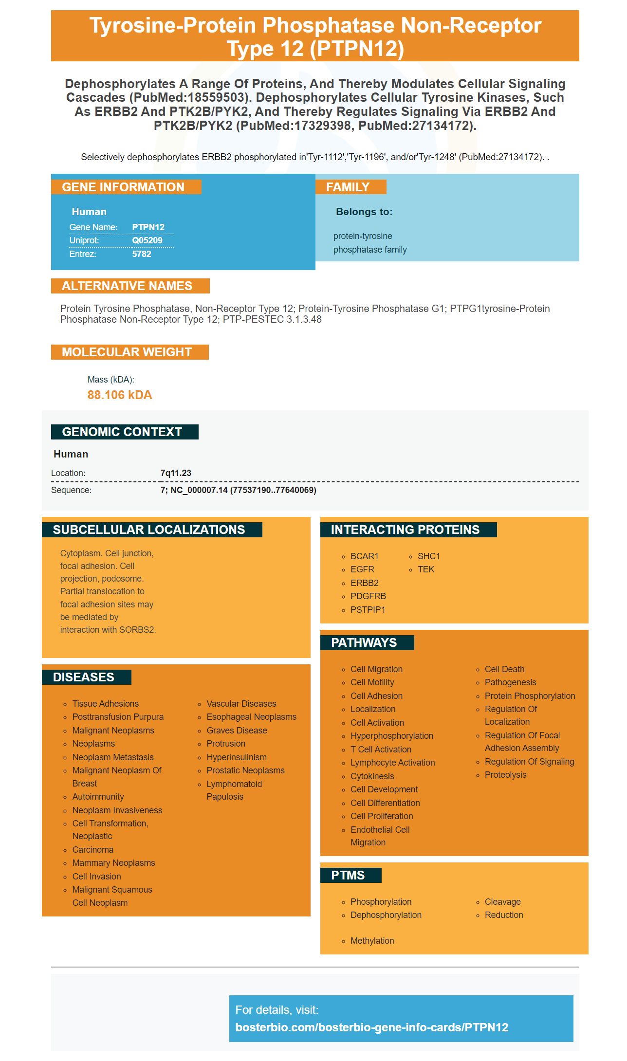
Facts about Tyrosine-protein phosphatase non-receptor type 12.
Selectively dephosphorylates ERBB2 phosphorylated in'Tyr-1112','Tyr-1196', and/or'Tyr-1248' (PubMed:27134172). .
Human | |
---|---|
Gene Name: | PTPN12 |
Uniprot: | Q05209 |
Entrez: | 5782 |
Belongs to: |
---|
protein-tyrosine phosphatase family |
protein tyrosine phosphatase, non-receptor type 12; Protein-tyrosine phosphatase G1; PTPG1tyrosine-protein phosphatase non-receptor type 12; PTP-PESTEC 3.1.3.48
Mass (kDA):
88.106 kDA
Human | |
---|---|
Location: | 7q11.23 |
Sequence: | 7; NC_000007.14 (77537190..77640069) |
Cytoplasm. Cell junction, focal adhesion. Cell projection, podosome. Partial translocation to focal adhesion sites may be mediated by interaction with SORBS2.
In this article, we will examine the Anti-PTPN12 PTPN12 Antibody PicoBand(tm) and discuss Boster Bio's process of product validation. Boster Bio's antibodies are validated on multiple platforms and with known positive and negative samples. We will discuss the various benefits of using Boster's PTPN12 antibodies and their applications.
The Boster Bio Anti-PTPN12 PTPN12 Protein Antibody PicoBand(tm), catalog #A02762-1, reacts with human PTPN12 recombinant protein. This product is stable at -20 degrees Celsius and 4 degrees Celsius, and contains 4 mg of Trehalose. A blocking peptide, available separately, can be used to block PTPN12.
Anti-PTPN12 antibody is a boster product with the catalog number A02762. This antibody reacts with Human and Mouse cells and is stable at -20degC for up to a year and can be stored at 4degC for up to a month. It comes in a liquid form in PBS containing 50 percent glycerol and 0.02% sodium azide. Boster products are validated for immunofluorescence, Western Blotting, and Immunohistochemistry.
This research demonstrates that alternative splicing results in distinct isotypes of PTPN12. These isoforms are potential biomarkers of cancer. Instead of quantification, they can be compared for detectability. This means that alternative splicing can provide a new way to detect cancer. Moreover, it can also help to identify potential biomarkers by using different peptides instead of a single peptide.
Although RNA splicing was first discovered in the 1970s, its significance was not fully appreciated until the Human Genome Project revealed that there are approximately 22,000 protein-coding genes in the human genome, and about 90,000 different proteins. Those studies laid the foundation for subsequent research, including the Human Proteome Project. In addition, alternative splicing has shown promise in early disease diagnosis.
Although this research is still in its early stages, these findings point to an important biological role for alternative splicing. As a result of alternative splicing, the expression levels of the corresponding proteins vary among individuals. Because alternative splicing involves the removal of non-coding regions, several proteins are encoded from a single gene. Moreover, alternative splicing facilitates gene regulation and increases the complexity of the proteome.
The authors concluded that alternative splicing results in distinct isotypes of PTPN12, a protein expressed primarily in the early stages of flower development, may play a role in flower development. To test the hypothesis, they sequenced 1716 alternatively spliced transcripts from three stages in flower development. These were further classified into four groups by a hierarchical clustering algorithm that considered all three stages simultaneously. The analysis revealed that 526 isoforms had higher expression levels in the IM stage compared to those of F1-9 and F12 stages, respectively.
Application of the PTPN12 marker is important for a variety of different purposes. This gene promotes cell survival in Gln-free conditions, and PTPN12 knockdown can prevent the apoptosis of MDA-MB-231 cells after 72 h of treatment. The PTPN12 gene is expressed in many human tissues and cells. Using a clone of ptpn12 knockdown cells, we can detect whether or not apoptosis has occurred in Ptpn12-KO cells after 72 h of treatment. We confirmed viability by using trypan blue exclusion.
Expression of PTPN12 is inversely correlated with poor prognosis in hepatocellular carcinoma, where loss of PTPN12 promotes tumor progression. PTPN12 is involved in regulating growth factor receptor dimerization and cytoskeletal reorganization. The loss of PTPN12 inhibits the activity of multiple oncogenic tyrosine kinases, including EGFR.
Expression levels of PTPN12 were measured using tissue microarray immunohistochemistry in two independent pathologists. The independent pathologists assessed PTPN12 expression in tumor tissue using a chi-square test. Expression levels were found to be associated with tumor stage and T and N classifications, but were not significantly correlated with age or sex. The authors conclude that PTPN12 is a valuable biomarker in the clinical setting for NPC.
Interestingly, shPTPN12 is involved in the modulation of the intracellular tumor suppressor CD99. Both p53 and PTPN12 are known to modulate the function of one another, although the exact relationship between them is still unknown. The PTPN12 gene is expressed on both sides of the cell membrane, and the cells of each side of the tumor display identical expression levels of PTPN12.
This study evaluated the prognostic impact of the PTPN12 marker in prostate cancer. This marker was highly expressed in cancers with ERG-negative status. Its prognostic value was found to be independent in three of the four scenarios analyzed. In contrast, PTPN12 expression was low or absent in ninety-four percent of ERG-positive tumors. Furthermore, PTPN12 expression was significantly associated with the presence of atypical cytogenetic marker, MMP-9.
In this study, cells were electroporated with three different crRNA against PTPN12. The cells were seeded in six wells and were incubated for two, three, or four weeks. Untreated cells were used as control samples. Non-targeting scramble crRNA complexes were used as a control. The data were collected from three independent experiments and presented as boxplots. Boxplots are interquartile ranges, while whiskers indicate the range from the minimum to maximum values. The median is shown as a line.
The PTPN12 marker was validated by assessing its sensitivity in breast cancer cell lines. In this study, a high PTPN12 staining score was associated with a good prognosis in PTEN-deficient cancers. Similarly, high PTPN12 expression correlated positively with a poor prognosis in Gleason 3 + 4 tumors. In addition, the PTPN12 marker was found to be highly expressed in tumors that had been previously classified as Gleason 3+4 cancers.
In a separate study, the PTPN12 marker was associated with poor survival in patients with hepatocellular carcinoma. This is consistent with the findings from the two previous studies. Furthermore, tumor-derived mutations affected the spatial configuration of the catalytic cleft, which likely inhibited the enzyme's function. Thus, further study on patients with hepatitis C is needed to validate the PTPN12 marker and confirm its utility for use in hepatocellular carcinoma.
PMID: 1472029 by Takekawa M., et al. Cloning and characterization of a human cDNA encoding a novel putative cytoplasmic protein-tyrosine-phosphatase.
PMID: 8454633 by Yang Q., et al. Cloning and expression of PTP-PEST. A novel, human, nontransmembrane protein tyrosine phosphatase.