This website uses cookies to ensure you get the best experience on our website.
- Table of Contents
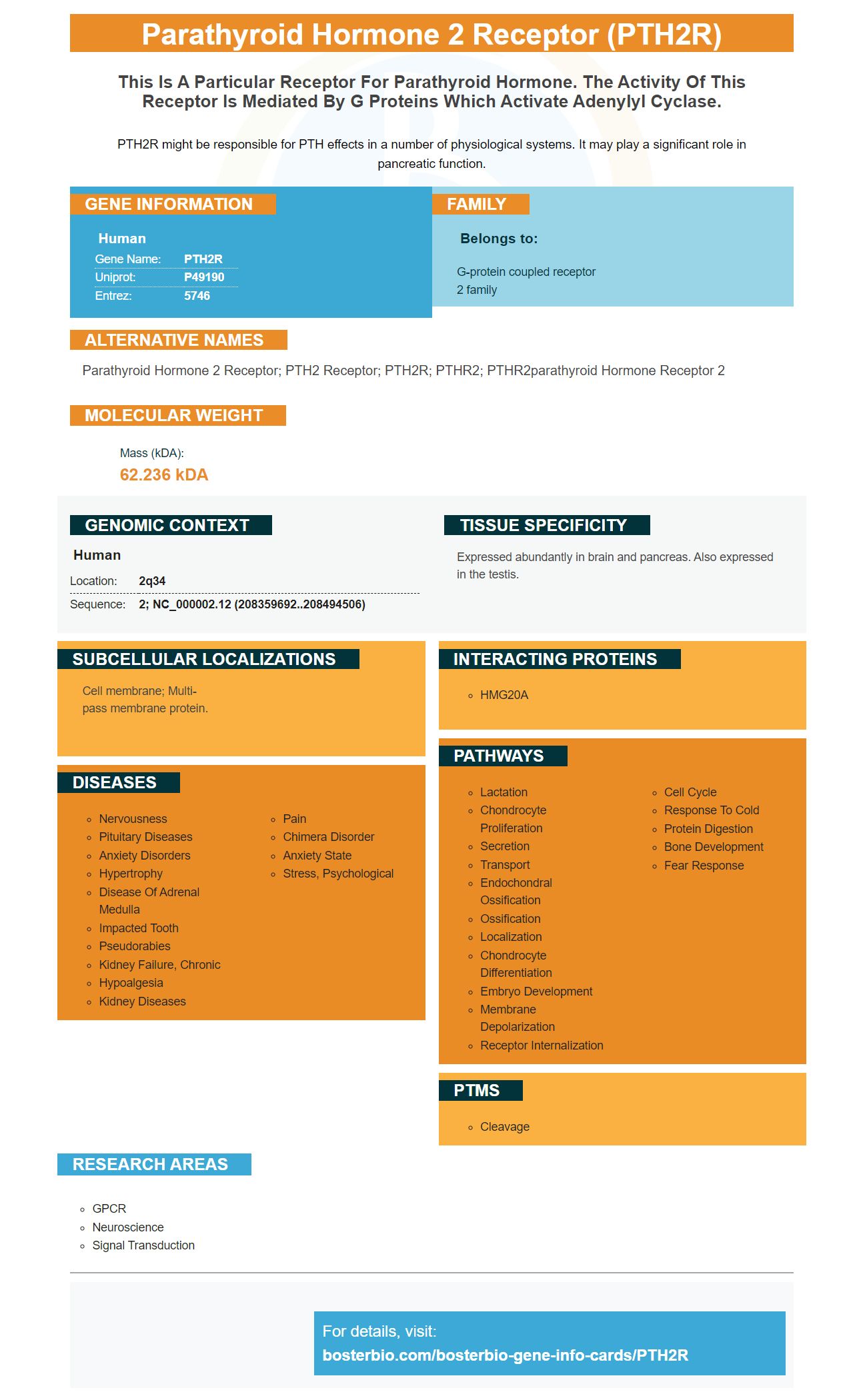
Facts about Parathyroid hormone 2 receptor.
PTH2R might be responsible for PTH effects in a number of physiological systems. It may play a significant role in pancreatic function.
Human | |
---|---|
Gene Name: | PTH2R |
Uniprot: | P49190 |
Entrez: | 5746 |
Belongs to: |
---|
G-protein coupled receptor 2 family |
parathyroid hormone 2 receptor; PTH2 receptor; PTH2R; PTHR2; PTHR2parathyroid hormone receptor 2
Mass (kDA):
62.236 kDA
Human | |
---|---|
Location: | 2q34 |
Sequence: | 2; NC_000002.12 (208359692..208494506) |
Expressed abundantly in brain and pancreas. Also expressed in the testis.
Cell membrane; Multi-pass membrane protein.
Scientists have a myriad of options for the PTH2R Marker. It has been found to block RUNX2 and increase Wnt/b–catenin signaling pathways. This makes it a desirable method for bone regeneration in both aged and diabetic subjects. Here are some of the most effective uses of PTHrP. These benefits apply to scientists around the world.
The study found that the drug may reduce the expression of RUNX2 and a protein called B-catenin in mice. After six weeks of administration, PTH 1-34 treatment resulted in a reduction in RUNX2 expression by as much as 19 percent and 46%, respectively. In contrast, the NS-treated control rats showed an increase in the expression of RUNX2 by only 0.01 percent.
These findings suggest that PTH and the Wnt/b–catenanin pathways are in synergy to aid in the healing of fractures in older mice. The Wnt/b–catenin signaling pathways could improve fracture healing and encourage osteoblast development. PTH inhibitors of the signaling pathway could also improve microcirculation and promote early healing of fractures.
Similar results were observed for Boster Bio PTH 1-34. The treatment increased b-catenin expression but not RUNX2. This suggests that PTHrP might suppress type X collagen by the PKA signaling pathway. In the future, PTHrP may be a candidate for blocking the Wnt/b-catenin pathway in mice.
Other than PTH, FGF23 and A-klotho are other cytokinesins which regulate bone metabolism. FGF23 is an indirect inhibitor of Wnt. It affects both the kidney and the parathyroid gland. As a result, Boster Bio PTH 1-34 could have the potential to work as a local osteo-inducing agent. The plasmin substrate-linker allows the endogenous plasmin molecule to be cleaved. Once activated, the protein is released into the tissues around it and enhances bone formation.
The positive effects of PTH were noted in the adult mice. Both PTH and CA mice showed an increase in bone-forming expression of b-catenin. However, osteoclast and Wnt/b-catenin-mediated bone formation are negatively regulated by DKKs and sclerostin. PTH inhibits both these proteins, however it is possible that it is due to the Wnt/b/catenin pathway. To find out if PTH influences bone growth, more research is required.
PTH was subcutaneously administered to mice during this study for two or six weeks. Bones were examined for histological features, including cartilage degeneration. Immunohistochemistry identified the expression of PTH2 receptor, sclerostin and dickkopf Wnt signaling pathway inhibitor-1. Additionally, PTH2R and B-catenin. Also, RT-qPCR was used to determine the amount of mRNA related to bone tissue.
A recent study has shown that PTH 1-34 can inhibit the differentiation of human articular the chondrocytes. PTH 1-34 was given to mice and rats suffering from azacytidine-induced OA. These results suggest that PTH may be able to reduce the activity of RUNX2, though we don't know the exact function of this hormone.
A recent study found that PTH 1-34 boosts the activity of b-catenin in bone, however, it did not significantly increase the level of protein of RUNX2. The study suggests that PTH could affect RUNX2 through a different route. To determine the effects of PTH 1-34 on bone development, researchers are currently evaluating it in animal models. Boster Bio also demonstrated that the hormone may increase osteoblast activity.
Researchers also found that adult CA mice expressed more MMP9, VEGF and OCN than WT mice. These changes were not statistically significant. However adult PTH mice had higher RUNX2 expression. While this result is encouraging but further research is required to confirm this conclusion. The researchers believe that this hormone can aid in slowing the aging process by controlling the expression of RUNX2.
The study also indicated that PTH could inhibit the expression of RUNX2. PTH reduced the expression of RUNX2 by one-fourths. This resulted in a reduction of nine and 19 percent respectively. In addition, it reduced the expression of b-catenin, sclerostin, and safranin-O. It also increased the expression of PTH2R (PTH2R), PTH2R and C-catenin in rats.
The study found that a medication that was linked to the Wnt/bcatenin pathway of signaling promoted fracture healing in aged mice as opposed to mice treated with PTH. The b-catenin pathway boosted osteogenic differentiation and proliferation of mesenchymal stem cell cells in the aged mice. The osteoclasts that were induced by PTH also showed better healing of fractures.
Antibodies against PTH 1-34 were used to study the effect of the hormone in bone formation. The antibodies react with mouse and human as well as rat. Additionally, Boster Bio Anti-RUNX2 antibody is able to bind to the RUNX2 protein. This study has some limitations. The antibody doesn't work on mice treated with PTH. As with other antibodies, it reacts with human cells in WB tests.
In addition to targeting osteoblasts, PTHrP may also stimulate angiogenesis, which can improve bone regeneration in aged and diabetic patients. It works by activating the Akt/Erk1/2 pathway, which focuses on fibroblasts and endothelial cell. This makes PTHrP's entry into bone marrow an attractive strategy to boost the growth of bone in diabetic and older patients.
Researchers examined wound healing in diabetic rats treated with PTHrP-2@CaAlg and HUVECs. As measured by CD31 immunohistochemical staining, wound healing rate in the treatment group was more. Patients treated with PTHrP had higher numbers of blood vessels.
PTHrP was also demonstrated to reduce the rate of osteoblast eruptions in the same study. It also augmented bone formation and Sharpey fiber density. The results revealed that PTHrP-treated SHEDs were more effective in their ability to regenerate bone tissue than mice treated untreated. In both cases PTHrP treatment reversed osteoporotic bone disease.
Recent research has shown that HUVECs and HFF-1 cells cultivates were grown with PTHrP-2. These cells were cultured in serum-free media and the addition of PTHrP-2. After 48 hours, the cells were harvested , and the supernatants were purified using a 0.22-mm filter. The treatment had a significant impact on cell proliferation.
The regulatory protein PTHrP is involved in osteoblast differentiation. PTHrP boosts the activity of both osteoblast-specific genes, including VEGF and BMP-2. It also inhibits alkalinephosphatase. In addition, PTHrP can reduce the expression of genes that are associated with terminal differentiation.
Sciencell Research Laboratories provided the cells used in this study. Cultures were performed in serum-free DMEM supplemented with 2.5% FBS, 1% ECGS, and penicillin-streptomycin. Only early-stage HUVECs were used in future studies. HFF-1 cells were grown with serum-free DMEM.
Exosomes are highly efficient in healing wounds. PTHrP-2 interferred exosomes increase the ability of endothelial cells to secrete collagen I. This protein is a key factor in HaCaT migration. PTHrP also improves the function of Akt/Erk1/2 in skin tissue.
For research into craniofacial stem cells PTH2R is a marker that can be extremely useful. To aid in the eruption of teeth, it's crucial to regulate PTH2R activity during its development. It also regulates osteogenic differentiation in OMSCs and play a role in the driving force behind the process of eruption. Here are some advantages of the PTH2R marker.
The PTH2R gene plays a crucial role in the metabolism of bone, calcium homeostasis and bone metabolism. Genetic studies suggest that the PTH2R gene's polymorphisms are essential markers for the regulation of bone turnover in Saudi Arabian women postmenopausal women. In addition, these gene variants are associated with the levels of bone turnover markers that are present in the bloodstream. This means that the PTH2R marker can be useful in identifying patients at risk of osteoporosis.
In addition, it can assist in identifying cancerous cells with different expression levels. In this instance those with high levels of the PTHrP marker would have a better chance of survival. A lower risk of lung cancer is associated with PTH2R-positive plasma cells. The PTHrP marker can also be used to differentiate between benign and malignant neuroblastic cancers.
The PTH2R gene encodes a seven-transmembrane receptor. The hormone plays an important function in a variety of physiological functions such as bone metabolism and mineral ion homeostasis. It also aids in the development of the skeletal structure. PTH2R is most frequently expressed in bones and kidney. PTH2R can also be useful in diagnosing osteoporosis or other bone disorders.
The PTH2R is a highly specific marker for calcium ions. The PTH2R signaling pathway is highly regulated by calcium ions. Calcium ions encourage interactions between PTH2R and spep. These interactions enhance the activity of the cAMP signaling. However, the essential mechanism of PTH2R allosterism is unknown. A large number of MD simulations were carried out to determine the role of calcium ions in the PTH2R/G-protein coupling. The simulations reveal that calcium ions increase receptor-G protein coupling.
PMID: 7797535 by Usdin T.B., et al. Identification and functional expression of a receptor selectively recognizing parathyroid hormone, the PTH2 receptor.
PMID: 8921382 by Usdin T.B., et al. Assignment of the human PTH2 receptor gene (PTHR2) to chromosome 2q33 by fluorescence in situ hybridization.