This website uses cookies to ensure you get the best experience on our website.
- Table of Contents
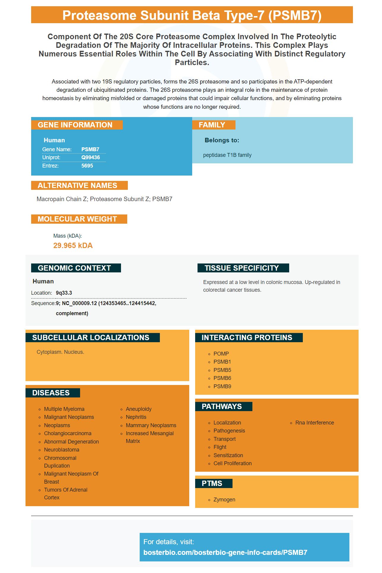
Facts about Proteasome subunit beta type-7.
Associated with two 19S regulatory particles, forms the 26S proteasome and so participates in the ATP-dependent degradation of ubiquitinated proteins. The 26S proteasome plays an integral role in the maintenance of protein homeostasis by eliminating misfolded or damaged proteins that could impair cellular functions, and by eliminating proteins whose functions are no longer required.
Human | |
---|---|
Gene Name: | PSMB7 |
Uniprot: | Q99436 |
Entrez: | 5695 |
Belongs to: |
---|
peptidase T1B family |
Macropain Chain Z; Proteasome Subunit Z; PSMB7
Mass (kDA):
29.965 kDA
Human | |
---|---|
Location: | 9q33.3 |
Sequence: | 9; NC_000009.12 (124353465..124415442, complement) |
Expressed at a low level in colonic mucosa. Up-regulated in colorectal cancer tissues.
Cytoplasm. Nucleus.
The PSMB7 indicator is made from mouse or rabbit. This antibody can be used to detect the protein on a wide variety of biomedical instruments, including Western blot, Autoradiography film, and immunohistochemistry. Scientists from all over the world are eligible to participate in the Boster Rewards program. It rewards researchers who discover new uses of the PSMB7 proteins. Here are some tips to make the most of the PSMB7 antibody:
Boster Bio has a high-affinity PSMB7 antigen (catalog number: A08095) for your research. Boster antibodies have a long history of validation on immunohistochemistry, Western Blotting, and ELISA. They are highly regarded by the scientific world. The following host cells are compatible with Boster PSMB7 antibodies: Rat, Mouse, and Human.
A PSMB7 monoclonal antibody is one that is produced by immunizing animals in the central region of human PSMB7 with synthetic Peptides. It recognizes both mature as well as precursor forms of the protein. This antibody does not cross-react, and it is compatible with PSMB10 (and PSMB5). The antibody was created in mice and rats and is suitable only for research. It should not be used in diagnostic procedures.
The PSMB7 gene is expressed in breast cancers, in which high levels of this protein are associated with poor prognosis. Inhibition of PSMB7 expression causes myogenic defects. PSMB7 antibody can be produced in rabbits or mice. These animals are good for research as they can tolerate high doses. However, they do not display a specific antigen. If you are interested and able to create PSMB7 antibody, make sure that you read the entire article.
The antisera were developed using myoblasts, 12h differentiating C3 cells, and control cells. The primary antibody consisted of a polyclonal, antiserum to PSMB7. The secondary antibodies were obtained by a LSAB+-HRP kit. They were used both in parallel and separately. We also performed a t-test of cell cultures on mice-derived cells.
In addition to developing PSMB7 antibodies, scientists used a transgenic mouse strain called Tg229 to test their antigen. The antibody was created from an immunized strain of mouse and was used to screen the protein in skeletal muscles cells. These antigens were injected into mice for several days. This study also produced antibodies against other subunits of skeletal muscle.
Eight exons make the PSMB7 genome. The plasmids required to produce PSMB7 antibodies contain puromycin resistant genes. These plasmids are created using the SureSilencing SHRNA gene from Qiagen. The inserted sequences were TCCGGAAGCTCCTAGGTAAT and ATGCTAATTCGACAGCCTAAT. The cells were then transfected using the SureSilencing shRNA virus using the Invitrogen Neon transfection system.
Western blots using the PSmb7 markers are widely used. This process involves separating the proteins according to their size and then transferring them to the membrane. To analyze the bands, primary antibodies are used, which recognize specific proteins. Each band should correspond to a target protein's molecular mass, which can then be determined using a protein ladder. Different methods have been refined over the years.
Different methods of Western blot detection use different antibody to identify specific proteins. One method uses a secondary antibody, which provides specificity to the protein of interest. After the secondary antigen is applied to a membrane, a specific substance solution is added. Photons are released when the enzyme reacts with secondary antibody. The light emitted in the reaction is then detected.
Fluorescent westernblot detection is a better option than chemiluminescent substrate detectors. For one, it is sensitive and preserves the sample. Second, fluorescent multiplexing provides a clear distinction between multiple targets. Composite images can be created that show single-color signals from individual proteins. A researcher can identify specific details by visually inspecting the signals one at a time. A fluorescent western blot is a great way to determine which proteins are interacted with which molecules.
There are several Western Blot detection methods that can be used to detect the presence PSMB7 proteins. PSmb7 has a high expression level in breast cancer and colon. This protein has been suggested to be a prognostic indicator for breast cancer. These two approaches can help researchers to better understand the biology of cancer. Western blot detection with the PSmb7 marker has many other benefits.
Both methods require a secondary antigen conjugated to an enzyme for quantitative analysis. AP has a narrower range of substrates, while HRP has a wider selection. And AP can be used to detect the PSmb7 protein. This technique uses an enzyme to create a colored product. However, colorimetric detection methods are more sensitive and require a high amount of protein.
PSMB7 is a part of the 20S core proteasome complex, which plays a number of essential functions in cells. It is associated with distinct regulatory particles and is detected by antibodies. Boster Bio has developed this antibody. It binds PSMB7 in many animal specimens. This antibody can be used in a variety of tissues including rabbit, mouse, and even human. It is used in many biological assays.
PMID: 8666937 by Hisamatsu H., et al. Newly identified pair of proteasomal subunits regulated reciprocally by interferon gamma.
PMID: 8610016 by Groettrup M., et al. A role for the proteasome regulator PA28alpha in antigen presentation.