This website uses cookies to ensure you get the best experience on our website.
- Table of Contents
8 Citations 6 Q&As
8 Citations 1 Q&As
5 Citations
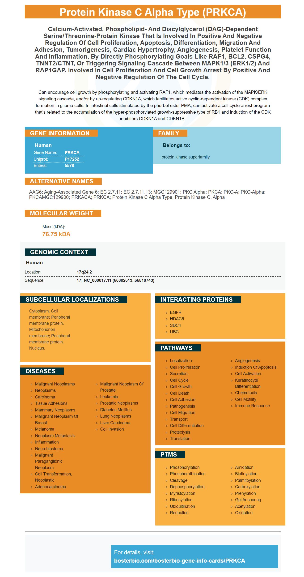
Facts about Protein kinase C alpha type.
Can encourage cell growth by phosphorylating and activating RAF1, which mediates the activation of the MAPK/ERK signaling cascade, and/or by up-regulating CDKN1A, which facilitates active cyclin-dependent kinase (CDK) complex formation in glioma cells. In intestinal cells stimulated by the phorbol ester PMA, can activate a cell cycle arrest program that's related to the accumulation of the hyper-phosphorylated growth-suppressive type of RB1 and induction of the CDK inhibitors CDKN1A and CDKN1B.
Human | |
---|---|
Gene Name: | PRKCA |
Uniprot: | P17252 |
Entrez: | 5578 |
Belongs to: |
---|
protein kinase superfamily |
AAG6; aging-associated gene 6; EC 2.7.11; EC 2.7.11.13; MGC129901; PKC alpha; PKCA; PKC-A; PKC-alpha; PKCAMGC129900; PRKACA; PRKCA; protein kinase C alpha type; protein kinase C, alpha
Mass (kDA):
76.75 kDA
Human | |
---|---|
Location: | 17q24.2 |
Sequence: | 17; NC_000017.11 (66302613..66810743) |
Cytoplasm. Cell membrane; Peripheral membrane protein. Mitochondrion membrane; Peripheral membrane protein. Nucleus.
We will be discussing the benefits of the PRKCA Marker as well as its biological functions in this article. We will also talk about its applications in research. This marker is extremely beneficial for scientists and researchers. Here are some examples:
A new study suggests that those affected by the PRKCA gene have increased risk of suffering from heart failure. The PRKCA gene is found in two variants that are common, is an excellent candidate for use in screening for heart failure. Studies have shown that this gene is linked to the shorter QT interval or longer QT interval. This is a risk factor for developing heart failure. Researchers have examined the relationship between this gene and other heart failure characteristics, such as LV wall thickness and fractional shortening, and have come to an agreement.
The rate of survival of various cancers is related to the expression of PRKCA genes. Its expression levels were significantly connected to survival rates in KICH and LIHC patients. It also correlated with survival in patients suffering from BRCA, LGG, MESO, and LUAD cancers. A PRKCA gene expression test can provide many advantages. It can be used to identify cancers with a poor prognosis.
Multiprotein complex that regulates cell motility and differentiation, the PKCa gene. It has been linked to cancer and positively or negatively related to tumor grade. It is also linked to breast cancer, and an inherited PRKCA gene variant is associated with an increased risk of PTSD in survivors of genocide. It is comprised of 672 amino acid and has a molecular value of 102 kD.
The PRKCA gene belongs to the PKC family. This gene is involved in a variety of cellular functions, including regulation of the ERK/MAPK pathway and PI3K/AKT pathway. It has been discovered to be highly upregulated in breast cancer colon carcinoma, NSCLC cell lines, and Hematological malignancies. Lahn and coworkers, who are from the University of California San Diego, discovered that the expression of the PRKCA gene was linked with different types of cancer.
Patients suffering from ACC, CESC and LGG had higher survival rates when they had high levels of the PRKCA marker. However, low levels of the PRKCH gene were associated with a poorer prognosis in UCEC and SKCM. However, PRKCQ, PRKCZ levels were positively related to survival in patients suffering from BRCA or LGG.
The expression of PRKCA was discovered to be associated with cancer recurrence after the last follow-up. Patients with higher PRKCA levels had a lower overall survival rate and were at greater risk of developing cancer. It was also associated with a lower prognosis, and a lower DFS. Age and sex did not appear to influence the results of this research.
The expression of PRKCA was analysed in 123 samples from LADC and 64 normal lung tissues. Both samples had high and low levels of PRKCA protein expression. LADC however, showed significantly higher levels of PRKCA protein expression than the LSCC or LADC cells. These results are in line with earlier studies that have demonstrated the value of PRKCA in the prevention of lung cancer. Further research is required to confirm its role in the prevention of lung cancer.
The PRKCA gene encodes a distinct member of the family known as the protein-kinase. PKCs are enzymes which add the phosphate group and alter their functions. They have been studied in many species including drosophila and xenopus. The gene is involved in cardiac and hepatic function. But its role in the cardiovascular system isn't well understood.
Furthermore, PRKCA and HDGF are linked to a lower survival rate among lung cancer patients. These findings suggest that LADC patients with higher levels of HDGF mRNA and PRKCA proteins are less likely to survive. However these findings do not exclude the possibility that increased levels of HDGF and PRKCA might contribute to the progression of lung adenocarcinoma. This could be the reason why these two proteins can be linked in the development of lung cancer.
The PRKCA marker that is a pleiotropic location, is associated with both obesity as well as asthma. A linkage analysis of BMI revealed significant connections between PRKCA SNPs in a population and BMI. The PRKCA marker was also shown to have a significant impact on adipocyte differentiation in both males and females. However, further functional studies are needed to understand the underlying mechanisms of these associations.
Previous studies have shown that the PRKCA gene is overexpressed in a variety types of cancers, including lung cancer. MiR-203 is the regulator of this increased expression. However, most studies focused on cell lines and not on tissue biopsies. Patients with lung cancer who have high PRKCA expression are more likely to be in poor health. MiR-203 regulates its expression. The presence of high PRKCA expression is linked with poor prognosis and tumour recrudescence and AJCC clinical stage.
This study provides a molecular basis for the development inhibitors of HDGF/PRKCA. This study was supported by the National Natural Science Foundation of China grant, China Postdoctoral Science Foundation grant, and a foundation for applied fundamental research between Kunming Medical University (China) and Yunnan Province. The authors acknowledge the financial support provided by the Yunnan Province and Kunming Medical University.
The PRKCA protein has been detected in 123 samples of LADC cells as well as in 70 normal lung tissues. The PRKCA protein is a major expression in LADC cells, is located in the cytoplasm. It is also significantly more prevalent in lung epithelial cell lines. The majority of samples contained high levels of PRKCA protein. Analyzing the statistics revealed that these cells were more likely than normal lung tissue to develop cancer.
The PRKCA marker was also associated with T and N classification. Additionally, it was associated with lymph node metastasis as well as the AJCC clinical stage however, it did not show any association with age or gender. This study provided more evidence about the relationship between the expression of PRKCA and survival rates for breast cancer. However, more research is needed to understand the role played by this marker for cancer. In addition to clinical uses, PRKCA has been found to be a valuable biomarker to aid in the research of cancer.
In the past the past, cell sorting and microscopic methods were employed to identify different types of cells. They take a long time and could cause suboptimal annotations. It is important to choose the right marker for every type of cell and cell cluster in the sample. This can only be accomplished through a computational platform that uses a comprehensive cell marker database.
PMID: 2336401 by Finkenzeller G., et al. Sequence of human protein kinase C alpha.
PMID: 1714454 by McSwine-Kennick R.L., et al. Phorbol diester-induced alterations in the expression of protein kinase C isozymes and their mRNAs. Analysis in wild-type and phorbol diester-resistant HL-60 cell clones.
*More publications can be found for each product on its corresponding product page