This website uses cookies to ensure you get the best experience on our website.
- Table of Contents
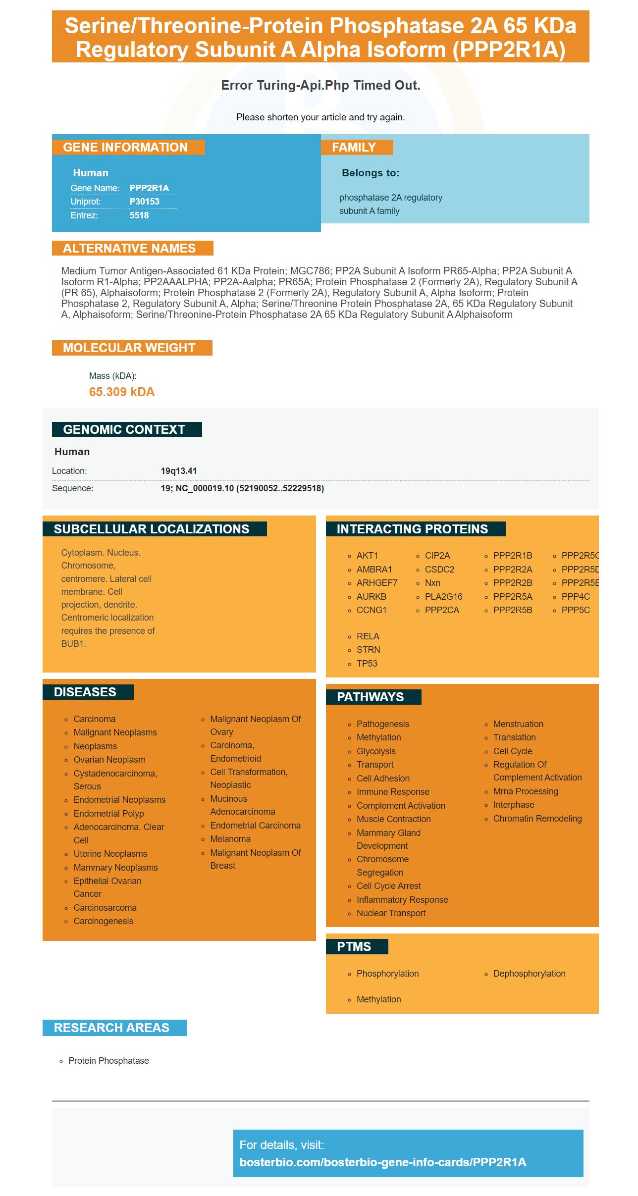
Facts about Serine/threonine-protein phosphatase 2A 65 kDa regulatory subunit A alpha isoform.
Please shorten your article and try again.
Human | |
---|---|
Gene Name: | PPP2R1A |
Uniprot: | P30153 |
Entrez: | 5518 |
Belongs to: |
---|
phosphatase 2A regulatory subunit A family |
Medium tumor antigen-associated 61 kDa protein; MGC786; PP2A subunit A isoform PR65-alpha; PP2A subunit A isoform R1-alpha; PP2AAALPHA; PP2A-Aalpha; PR65A; protein phosphatase 2 (formerly 2A), regulatory subunit A (PR 65), alphaisoform; protein phosphatase 2 (formerly 2A), regulatory subunit A, alpha isoform; protein phosphatase 2, regulatory subunit A, alpha; serine/threonine protein phosphatase 2A, 65 kDa regulatory subunit A, alphaisoform; serine/threonine-protein phosphatase 2A 65 kDa regulatory subunit A alphaisoform
Mass (kDA):
65.309 kDA
Human | |
---|---|
Location: | 19q13.41 |
Sequence: | 19; NC_000019.10 (52190052..52229518) |
Cytoplasm. Nucleus. Chromosome, centromere. Lateral cell membrane. Cell projection, dendrite. Centromeric localization requires the presence of BUB1.
There are many reasons to use this reagent. In this article, we will explore the most common applications. For example, you can use it to investigate the role of PPP2R1A in germ cell tumors. This reagent is also known as PLAP. PLAP is a protein produced by the placenta. The PPP2R1A gene is responsible for regulating the amount of PLAP in the placenta.
A recent study found that the expression levels of two genes are significantly higher in female placentas than in male placentas. These genes are both linked to the X chromosome and escape X-inactivation in humans. Hence, studying these genes in mice may have a number of benefits for human pregnancy. However, further studies are needed to determine whether they have any physiological or medical significance.
This study also highlighted the role of the Polycomb group gene L3mbtl2 in the development of the early labyrinth. Using a database of embryonic lethal mutants, this study discovered that the GPI-anchor biosynthesis pathway is a key player in the formation of the early labyrinth. Likewise, efforts to identify novel genes and subviable mutants are of great value in identifying functional protein complexes involved in normal placental development and the occurrence of IUGR.
The analysis of recent mouse mutants opened many new directions for research. It has shown that most embryonic lethal mutations result in placental defects. It also highlighted the possibility of novel molecular networks during placentation. Many of the genes involved in placental development have already been implicated in human placentation and developmental problems. This study is only the first to identify novel placental uses of the PPP2R1A marker.
As the placenta is the chief regulator of the nutrient supply to the growing embryo, its proper function is crucial for its proper development. Placental defects affecting the placenta can result in intrauterine growth restriction. Despite the similarities between human and murine placentas, human placentas are significantly more complex. Therefore, the precise diagnosis of placental malformation may be difficult.
The labyrinth layer is an important component of the placenta. It serves as an exchange surface for nutrient and gas exchange. Defects in the labyrinth cause intrauterine growth restriction (IUGR) and fetal lethality. In many cases, defects in the labyrinth are the result of defective invagination of allantoic blood vessels into the chorionic ectoderm. The development of the exchange surface requires branching morphogenesis.
The mouse labyrinth closely resembles the human placenta, although the exact equivalent is not known. However, it is thought that the cytotrophoblast cell columns (CC) are analogous to the JZ in human placentas. They anchor the placenta to the uterine wall, and are densely packed and highly proliferative. Extravillous cytotrophoblast cells invade the uterine stroma.
The serum tumour markers bHCG and bHCE are valuable tools in the clinical management of germ cell tumours (GCTs). Since they were first used in the 1970s, they have been widely used to aid in timely diagnosis, stage disease, monitor response to treatment, and detect relapse during follow-up. The marker is produced primarily by syncytiotrophoblastic giant cells (STGs) and chorionic carcinoma.
In addition, the antigen of Boster Bio is a recombinant protein that reacts to Human cells. In addition to detecting germ cell tumors, it can also identify the type and extent of cancer cells in a sample. To obtain accurate results, you must first understand which gene or protein in the tumor is causing the inflammation. Boster Bio has designed its antibodies to recognize and react with these genes.
The recombinant TSPY protein is expressed in seminomatous and nonseminomatous germ cell tumors. Furthermore, it could be a useful diagnostic marker for germ cell tumor precursors and help in the subtyping of TGCTs. The gene also plays an important role in subclassification and subtyping. In addition to these clinical benefits, TSPY expression can be used as a screening tool for other tumor types, including nonseminomas.
As of January 2018, there are three testicular cancer types that secrete TSPY: seminomas and nonseminomas. The other two types of tumors are mixed germ cell tumors (MGCTs), which are defined as having a mixture of GSTCs. While the TSPY gene is a candidate gene for GBY, it is still in early development and has not been confirmed.
Using a tumour marker is an easy way to diagnose germ cell tumours, as it can also be confirmed through scan results. It can also help doctors determine tumour stages. Stages refer to how big the tumour is and whether it has spread to other parts of the body. Secondary cancers are also diagnosed with a tumour marker and treatment is dependent on the stage and location. It can be treated through surgery or chemotherapy.
Intermediate germ cell tumors have several markers that may help in detecting them. These markers include MAGEA4, c-KIT, and NANOG. Intermediate germ cells also express markers that distinguish them from other types of germ cells, such as TSPY and VASA. In addition to these, they express markers associated with germ cell development. The marker also helps doctors detect germ cell tumors by analyzing their morphology.
Placental alkaline phosphatase, or PLAP, is an enzyme that hydrolyzes phosphoric acid monoesters. It is an oncofetal antigen and a member of a family of four alkaline phosphatase isoenzymes. Human germ cell tumors express PLAP and its related isoenzymes. Antibodies to PLAP react with syncytiotrophoblasts in the placenta and ovarian germ cell tumors in human.
Human PLAP was purchased from Sigma Aldrich and dissolved in a solution of 25 mM HEPES and 100 mM NaCl. This sample was filtered through a 0.22 um PES membrane and centrifuged in a Vivaspin(r) 20 MWCO 300,000 device. The purified PLAP was tested by size-exclusion chromatography for activity.
Placental alkaline phosphatase is expressed in the placenta during the third trimester of pregnancy. Anti-AP antibody identifies this enzyme in germ cells of the testis, endometrium, and testis. The anti-PLAP antibody does not cross-react with other alkaline phosphatases.
Placental alkaline phosphatase is related to ovarian cancer and has been shown to be a promising biomarker for early detection of ovarian cancer. Several fully human monoclonal antibodies to PLAP bind with a dissociation constant of 10 nM. In addition, immunofluorescence and Biacore analyses have shown that B10 antibody specifically localizes to tumors with A431 mutation. These antibodies may serve as a modular building block for targeted therapeutic products and radionuclides.
The PLAP monoclonal antibody has been developed by Santa Cruz Biotechnologies. This antibody is the 8B6 clone. It is available in a molar concentration of 10 ug/ml in PBS 1% BSA. For quantitative biodistribution experiments, radioiodinated SIP(B10) was injected into nude mice. Tumor:organ ratios ranged from two to 7.5.
As a tissue-specific antibody, anti-PLAP is specific for PLAP in human tissues. Its monomeric form is found in human placenta tissue. The placenta contains abundant levels of PLAP. The staining pattern of PLAP was comparable to that of the commercial anti-PLAP 8B6 murine antibody. The negative control for this assay was an anti-hen egg lysozyme KSF antibody and phosphate-buffered saline solution. Anti-PLAP antibodies B10 and D9 also recognized PLAP.
PMID: 2554323 by Walter G., et al. Molecular cloning and sequence of cDNA encoding polyoma medium tumor antigen-associated 61-kDa protein.
PMID: 2159327 by Hemmings B.A., et al. Alpha- and beta-forms of the 65-kDa subunit of protein phosphatase 2A have a similar 39 amino acid repeating structure.