This website uses cookies to ensure you get the best experience on our website.
- Table of Contents
2 Citations 4 Q&As
1 Citations 3 Q&As
1 Citations 4 Q&As
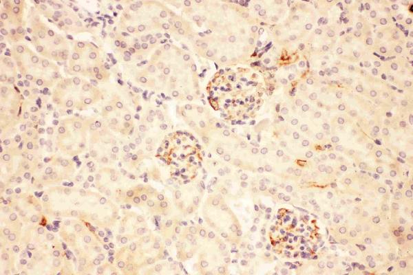
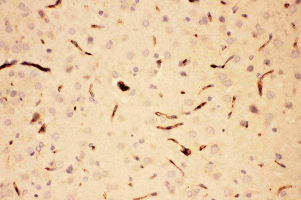
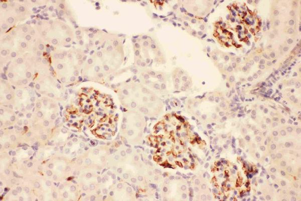
Facts about Pro-opiomelanocortin.
Human | |
---|---|
Gene Name: | POMC |
Uniprot: | P01189 |
Entrez: | 5443 |
Belongs to: |
---|
POMC family |
ACTH; adrenocorticotropic hormone; adrenocorticotropin; alpha-melanocyte-stimulating hormone; alpha-MSH; beta-endorphin; beta-LPH; beta-melanocyte-stimulating hormone; beta-MSH; CLIP; corticotropin-like intermediary peptide; corticotropin-lipotropin; gamma-LPH; gamma-MSH; lipotropin beta; lipotropin gamma; LPH; melanotropin alpha; melanotropin beta; melanotropin gamma; met-enkephalin; MSH; NPP; POC; POMC; pro-ACTH-endorphin; proopiomelanocortin preproprotein; proopiomelanocortin; pro-opiomelanocortin
Mass (kDA):
29.424 kDA
Human | |
---|---|
Location: | 2p23.3 |
Sequence: | 2; NC_000002.12 (25160853..25168851, complement) |
ACTH and MSH are produced by the pituitary gland.
Secreted. Melanocyte-stimulating hormone alpha and beta-endorphin are stored in separate granules in hypothalamic POMC neurons, suggesting that secretion may be under the control of different regulatory mechanisms.
The POMC marker is used in a variety of different areas of immunology. The POMC marker was recently employed to detect cancerous cells. POMCs are created by myeloid cells. They are found in many tissues. POMC is an uncommon marker due to its low expression in normal cell cells. This article will discuss the most common POMC markers and then discuss their uses. This article will also discuss the incubation of whole-cell extracts with antibodies that target the POMC.
RTN3 tagged with Xpress moves closer to the 25-kDa band of HEK-293 microsomes. This suggests that an anti-C-terminal antibody could induce RTN3 incorporation into the bilayer of lipids. The antibody can also detect the N-terminal tag Xpress and detect the R454 epitope, which is recognized by. The results from this study are in accordance with those of other groups that use RTN3 as the target protein.
Neuronal differentiation is associated with complex rearrangements of RTN3 expression. It is not known what RTN3 interacts the Bcl-2 family proteins. The survival of neurons and their plasticity are both affected by RTN3 and its variants. Incubation of whole cell extracts with RTN3 antibody is an important method for studying these proteins.
The antisera used for this experiment were specific for RTN3 (N-terminal fragment was pfu-polymerase-activated) and had high reproducibility. The immunoreactive bands that resulted were examined using the same antisera, which was pre-incubated using 100 mg of epitope. In all panels, mol mass was measured in kD.
The cells that were knocked down by siRTN3 showed decreased levels of RTN3. The RTN3 rescue cell line however, showed strong synthesis of viral proteins and replicative dsRNA. In addition, the immunofluorescent foci were significantly decreased in siRTN3 knockdown cells when compared with vector control cells and the parental. The results of these studies reveal that RTN3 knockdown cells do not block the replication of EV71, but the immune system is required to detect the protein.
The RTN3-enhanced growth of neurites was reduced by the reduction of protrudin. Additionally, the increased expression of RTN3 in DCs inhibited DC axon regeneration. This suggests that RTN3 is a potential treatment for SCI and may even help improve function in patients. Researchers are now looking into the possibility of developing RTN3 targeted therapies that are based on these findings.
Overexpression of RTN3 enhances the length of neurites in retinal cells. It also increases the transport of kinesin-1 which is linked to protrudin. Thus, it is believed that RTN3 overexpression may facilitate neurite outgrowth as well as axon regeneration. These findings could be relevant for neurodegenerative diseases, particularly in the retina. Incubation of whole-cell extracts using RTN3 antibody can help to detect these changes.
We took confluent cells from a tissue culture dish to find out if RTN3 was present. The pellets were then suspended in protease inhibitor mixtures. The cell homogenates were then centrifuged at 6800 rpm for 15 min. The supernatant was then transferred onto a centrifuge column at 4.degree Celsius for another 5 minutes.
In PV-induced vesicles RNA, DNA and viral proteins are synthesized. RNA moves to the ER where it forms complexes with products of translation. The ER is a replication factory and crystallization center. In RTN3 knockdown cells the rate of formation of replication complex-associated vesicles declines. The ER surface still has a residual complex. RTN3 knockdown does not affect viral protein synthesis , but it hinders the budding process and the creation of replication complex vesicles.
RTN3 overexpression promotes neurite outgrowth in naive DRGN cultures. Knockdown of RTN3 has no effect on the survival of DRGNs, but hinders the growth of neurites. This results suggests that RTN3 is responsible for neurite outgrowth. This is why whole-cell extracts infused with RTN3 antibodies aren't enough.
This study looked at the effects of pCMVFLAG-PGMCR on the morphology and morphology MP cells. Freshly-seeded WT cells showed a predominant elongated appearance with a few rounded cells. However, MP and DM cells had a large number of rounded cells. After 72 hours the proportion of cells that were round was significantly reduced in comparison to the WT cells. The plasmids that were used for these cultivars did not significantly alter the morphology of MP and DM cells.
PGRMC1 is located in the nucleus of bovine cells and oocytes. It is involved in the final meiosis in bovine-granulosa oocytes and bovine granulosa cell meiosis. Its involvement in drug binding, signaling and other aspects of its biology are well-known.
In this study, a pCMV-FLAG-PGMCR was co-transfected into HuH7 and HCT116 cells with Lipofectamine 2000 (Invitrogen). The cells were then for a night incubation with GL after transfection. The extracts were then lysed using NP40 the lysis buffer. To determine bound proteins, anti-FLAG agarose (60 min) was added to the lysates of cells. The bound proteins were then identified by western blotting using antibodies.
PGRMC1 could regulate the growth of tumors by reducing the amount of LDL in the cell. Therefore, inhibition of PGRMC1 may be beneficial to the growth of tumors. The inhibition of PGRMC1 can also alter the activities of genes that stimulate apoptosis and cell survival. These findings point to areas of research to be pursued. The results of these studies are positive.
Antibodies to PGRMC1 target the epitopes of the protein. PGRMC1-FLAG-PGRMC1-tagged proteins are made by using glutathione-S-transferase mutants. These mutations cause the PGRMC1 phenotypes. PGRMC1.
The cells were first kept at -80 degrees Celsius in Bambanker prior to the experiments. Then, the cells were returned to the laboratory in six mL of complete DMEM with Hygromycin B added to each well. After 48 h, cells were seeded threefold Dilutions in total DMEM.
After two million cells were resuspended an phenol-red-free DMEM/F12 culture medium A single-cell culture dish was employed. The medium was then diluted and then was supplemented with 10 Fetal Calf serum. One hundred and twenty mice were used for this study. The number of mice used was determined by the replicate coefficient of variation for solid tumor models. The coefficient of variation that replicates indicates that 10 mice are required to detect a 20% change.
As one would expect, PGRMC1 and DSUMO-PGRMC1 fusion proteins co-localize in the nucleus. Incredibly, P4 reduces the effects of pGRMC1 as well as DSUMO-PGRMC1-Flag. The P4 inhibitor also reduces Tcf/Lef activity by nearly twofold.
The effects of pCMV's pCMV-FLAG-PGM the human glioma cell morphology are not yet known. However, MAT can cause cell mobility and altered motility. Furthermore, the phosphorylation status of PGRMC1 regulates cell mobility. PGRMC1 phosphorylation status can affect mitosis.
Mass spectrometry was used to determine the function of GL in the heme-dimerized PGRMC1 and found that it was crucial. We found that PGRMC1 connects to GL and heme dimerization in two distinct regions of the protein. The L145-F162 residues show the most reduction in deuterium exchange with GL and F163-E173 amino acids showed the greatest increase.
PMID: 6274691 by Takahashi H., et al. Isolation and structural organization of the human corticotropin- beta-lipotropin precursor gene.
PMID: 6299668 by Whitfeld P.L., et al. The human pro-opiomelanocortin gene: organization, sequence, and interspersion with repetitive DNA.
*More publications can be found for each product on its corresponding product page