This website uses cookies to ensure you get the best experience on our website.
- Table of Contents
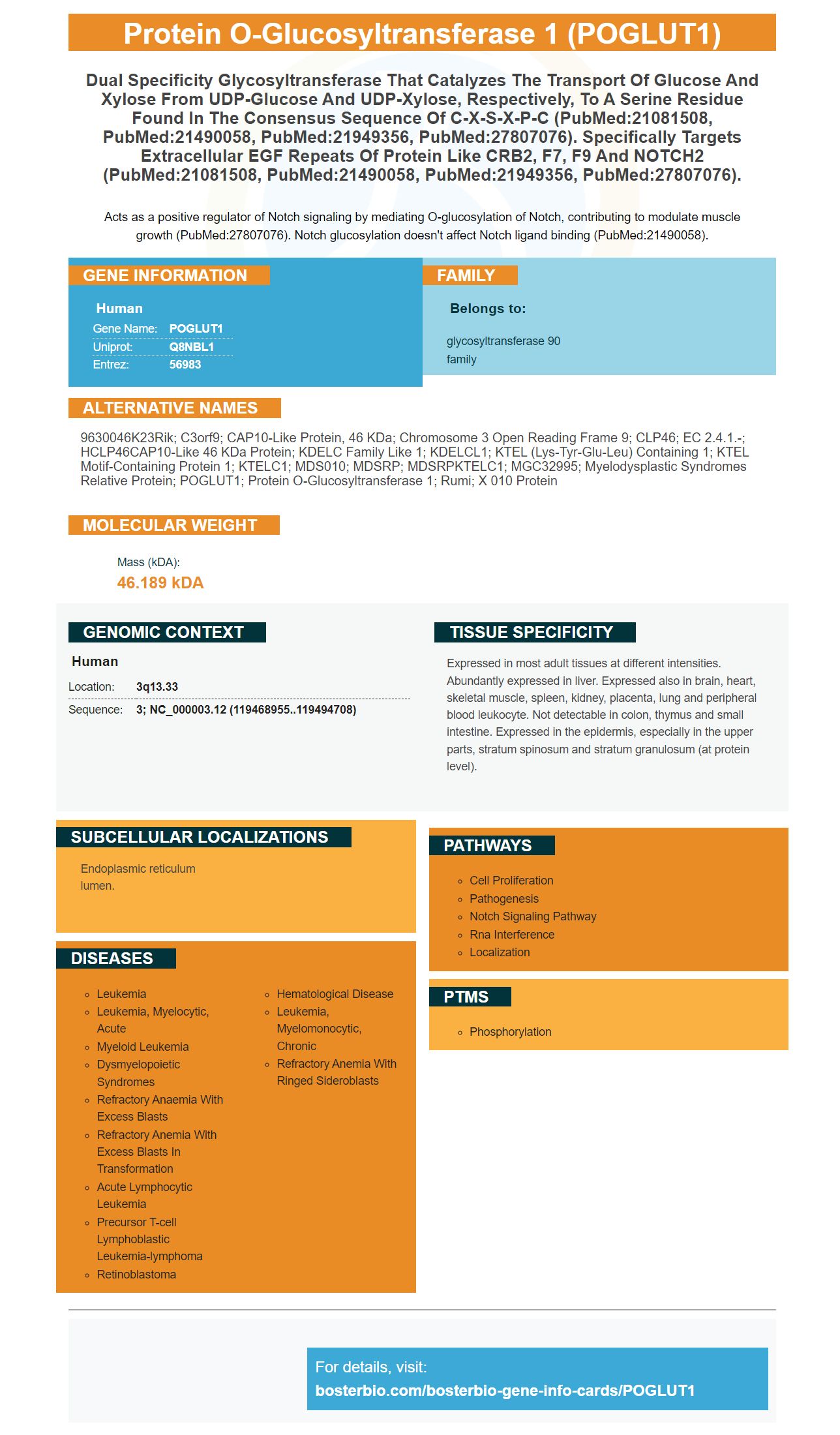
Facts about Protein O-glucosyltransferase 1.
Acts as a positive regulator of Notch signaling by mediating O-glucosylation of Notch, contributing to modulate muscle growth (PubMed:27807076). Notch glucosylation doesn't affect Notch ligand binding (PubMed:21490058).
Human | |
---|---|
Gene Name: | POGLUT1 |
Uniprot: | Q8NBL1 |
Entrez: | 56983 |
Belongs to: |
---|
glycosyltransferase 90 family |
9630046K23Rik; C3orf9; CAP10-like protein, 46 kDa; chromosome 3 open reading frame 9; CLP46; EC 2.4.1.-; hCLP46CAP10-like 46 kDa protein; KDELC family like 1; KDELCL1; KTEL (Lys-Tyr-Glu-Leu) containing 1; KTEL motif-containing protein 1; KTELC1; MDS010; MDSRP; MDSRPKTELC1; MGC32995; Myelodysplastic syndromes relative protein; POGLUT1; protein O-glucosyltransferase 1; Rumi; x 010 protein
Mass (kDA):
46.189 kDA
Human | |
---|---|
Location: | 3q13.33 |
Sequence: | 3; NC_000003.12 (119468955..119494708) |
Expressed in most adult tissues at different intensities. Abundantly expressed in liver. Expressed also in brain, heart, skeletal muscle, spleen, kidney, placenta, lung and peripheral blood leukocyte. Not detectable in colon, thymus and small intestine. Expressed in the epidermis, especially in the upper parts, stratum spinosum and stratum granulosum (at protein level).
Endoplasmic reticulum lumen.
Boster Bio optimization guidelines are one of the most effective ways to optimize your experiment. There are many options available for optimizing your experiment. This guide will help you record your test results using autoradiography film or ECL chemiluminescence. The following sections will help you optimize your experiments. Continue reading for more information about optimizing your experiments using the marker POGLUT1.
Detection of protein transfer efficiency by using membrane staining requires transferring a sample of proteins to a polyacrylamide gel. The size, abundance, charge, and hydrophobicity of proteins all affect their ability to transfer to the membrane. Because all proteins don't move at the same speed, it is impossible to transfer them all. After the transfer is completed, a small amount remains on the membrane.
A useful method to determine the transfer effectiveness of a protein is membrane staining with total protein stains. These stains can be very reliable while dyes can affect antibody binding. MemCode reversible cell stain is compatible with both nitrocellulose and PVDF membranes. Detection of protein transfer efficiency by membrane staining involves a gel that contains eight-cm-by-10-cm-size microgels.
The efficiency of protein transfer depends on the pore size of electroblotted membrane. A membrane with a diameter of 0.2mm is better for proteins below 20kD. A membrane with a molecular mass of 150kD will transfer a protein below 15 kD more easily to a polyacrylamide membrane.
The membrane staining method is used to determine the efficiency of protein transfers. This is crucial for quantitative western-blotting applications. While there are methods for loading a specific amount of protein onto the gel before electrophoresis, it is still difficult to measure the efficiency of transfer to nitrocellulose. Non-transfer, air bubbles, and other impurities can interfere with quantitative Western blotting. Fortunately, a standardized solution exists for this problem - StainODyeTM. You can see the bands of proteins on the transfer medium by using membrane staining. For permanent records, you can also take photos of stained membranes.
The use of membrane staining to detect protein transfer efficiency is a simple, easy method that requires very little preparation. It requires a solution that contains 1% KOH. There are several rinses with PBS. After incubation at 37°C for 30 minutes, the membrane should be rinsed with water several times. After the membrane has been thoroughly rinsed, it should be stained with Tween 20 solution. This solution also effectively stain the nitrocellulose membranes or PVDF membranes.
Autoradiography uses radioactive substances to take a photo of a patient's entire body and record the results. The film shows the irradiated regions as dark areas. The tracer distribution corresponds with their presence or absence. The process is widely utilized in radiology as well as medicine. Here are some benefits to using autoradiography films
In vitro Autoradiography allows you to see the anatomical distributions for a protein or other lipid. It involves the binding of radioligand to a specific target in frozen tissue sections. The radioligand can be detected by photosensitive film or phospho imaging plates. Digital autoradiograms allow for high spatial resolution, which allows for quantitative analysis on ligand affinity. This allows for diagnostics in a wide array of situations, including drug design.
The base of autoradiography films is made up of a thin layer clear polyester material. It provides support for all other components of the film. Although the base is not involved in the image-forming process of autoradiography film, some films are dyed lightblue to enhance the appearance of the image when viewed through a viewbox. The active component of the film, or emulsion is made up of small silver halide crystals suspended within gelatin. The emulsion is usually 10 um thick.
Autoradiography offers another advantage, namely the ability to study radioligand binding to tissue. This technique is especially useful in drug discovery and development programs. It provides valuable information about the distribution and pharmacokinetics of drugs. This is one way to best record test results. This technique is extremely reliable and can be used in clinical trials to identify promising candidates. You can use autoradiography to detect potential drugs in animals.
The use of enhanced luminol chemiluminescence (ECL) has many benefits over conventional western-blotting. ECL detects secondary proteins bound to primary antibody with high sensitivity, even when there is a low concentration. ECL substrates can be easily stripped and probed with various antibodies, if desired. It is a great choice for researchers trying to monitor the efficiency of protein transfers to cells.
In this method, proteins are separated based on their molecular mass, AA sequence, or primary structure. A protein sample is a mixture pre-defined proteins that confirms a particular mass band is correct. The protein standard is loaded in both the first and last lanes. The selection of a protein standard depends on the sensitivity of the gel, the resolution required near molecular mass, the size and analysis potential. Some protein standards come pre-stained to allow for easy visualization and to confirm the effectiveness of protein transport on membranes.
The signal intensity will depend on the amount and composition of the buffer components as well as the primary antibody. For proteins with a higher molecular mass, detergents as well as enzymatic inhibitors are necessary to improve their solubility. Negative and positive controls can be used to reduce background and nontarget binding. The linear dynamic range of the detection device should allow for wash concentrations and wash durations that are within the limits of the linear dynamic range. Quantification can either be done whole-lane or boxed. Other methods include Coomassie, rolling ball algorithm and boxed analyses. These methods can also serve to normalize results.
PMID: 16524674 by Teng Y., et al. Cloning, expression and characterization of a novel human CAP10-like gene hCLP46 from CD34+ stem/progenitor cells.
PMID: 21490058 by Fernandez-Valdivia R., et al. Regulation of mammalian Notch signaling and embryonic development by the protein O-glucosyltransferase Rumi.