This website uses cookies to ensure you get the best experience on our website.
- Table of Contents
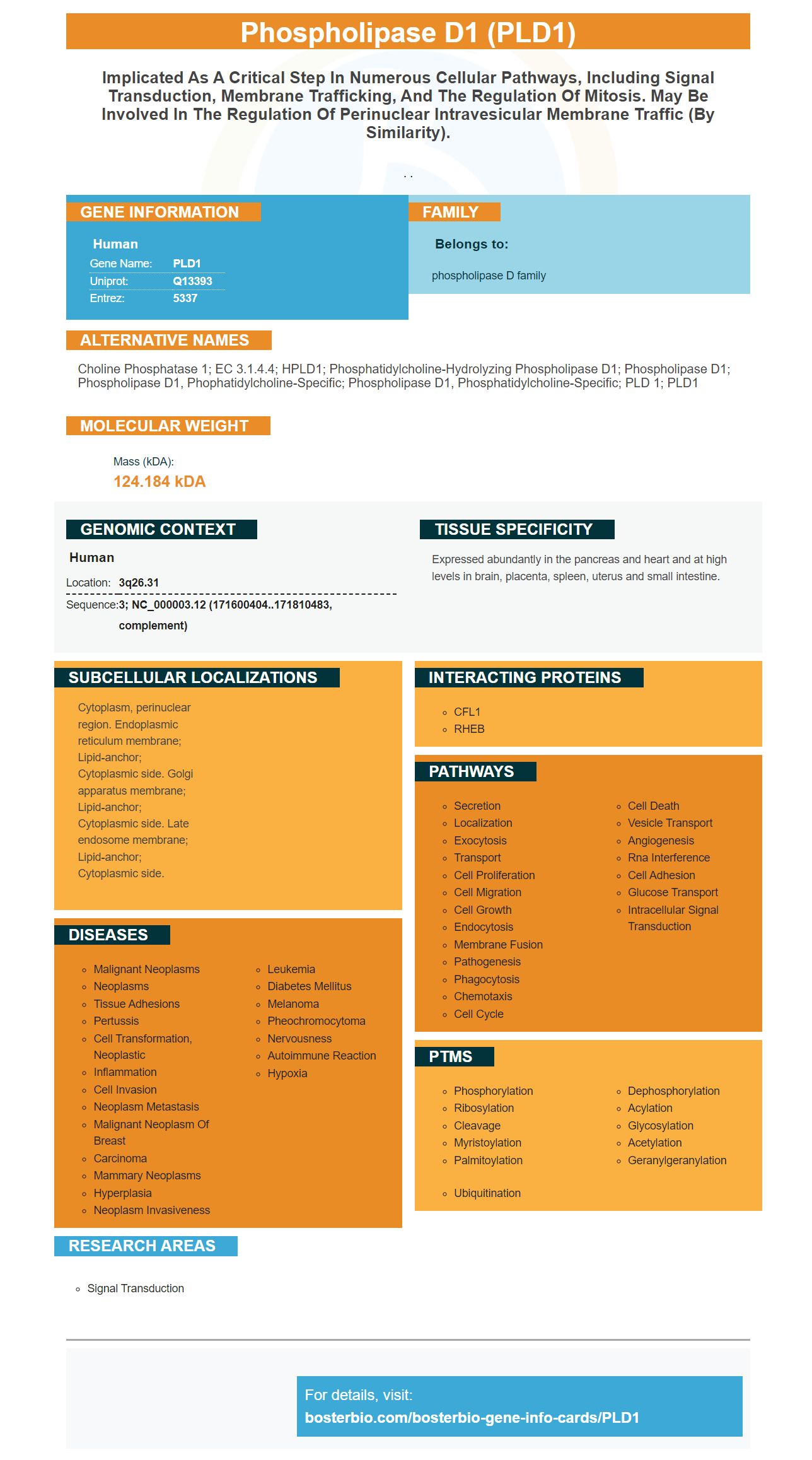

Facts about Phospholipase D1.

. .
| Human | |
|---|---|
| Gene Name: | PLD1 |
| Uniprot: | Q13393 |
| Entrez: | 5337 |

| Belongs to: |
|---|
| phospholipase D family |

Choline phosphatase 1; EC 3.1.4.4; hPLD1; Phosphatidylcholine-hydrolyzing phospholipase D1; phospholipase D1; phospholipase D1, phophatidylcholine-specific; phospholipase D1, phosphatidylcholine-specific; PLD 1; PLD1
Mass (kDA):
124.184 kDA

| Human | |
|---|---|
| Location: | 3q26.31 |
| Sequence: | 3; NC_000003.12 (171600404..171810483, complement) |
Expressed abundantly in the pancreas and heart and at high levels in brain, placenta, spleen, uterus and small intestine.
Cytoplasm, perinuclear region. Endoplasmic reticulum membrane; Lipid-anchor; Cytoplasmic side. Golgi apparatus membrane; Lipid-anchor; Cytoplasmic side. Late endosome membrane; Lipid-anchor; Cytoplasmic side.


If you are wondering how immunohistochemistry works, read on. Immunohistochemistry uses the interaction between an antigen and an antibody to identify proteins within tissues. By preparing the samples and staining them, researchers can visualize the distribution and localization of specific proteins in a variety of cell types. Successful immunohistochemistry results produce a strong signal and can be interpreted accurately.
The antibody used for IHC is a proprietary protein detection method that has been validated for use in the clinic. Its quality is unmatched with more than 16,000 antibodies validated for IHC, WB, ELISA, FC, and Western Blotting. The company offers mouse and rabbit polyclonal antibodies and a free secondary antibody for every purchase. The antibody itself is highly sensitive, and has been used for over 25 years in a variety of diagnostic applications.
The technique detects antigens or haptens within biological tissues using antibodies and enzymes. It takes advantage of the principle that antibodies bind to antigens and visualize their distribution in biological tissues. Boster Bio's guide to immunohistochemistry offers troubleshooting tips and techniques for common problems with IHC. It also provides a comprehensive guide to resources for IHC. Boster Bio has highlighted key IHC protocols and techniques for the purposes of diagnosis and research.
To confirm the specificity of an antibody, an absorption control is used. This antibody is incubated overnight with the immunogen to test the specificity of the primary antibody. Then, an inactivated antibody is used to replace the primary antibody in the IHC protocol. This ensures that the inactive antibody does not stain at antibody binding sites. Positive tissue controls, on the other hand, should not have any expression of the target antigen.
Another popular immunohistochemistry method is ELISA. Using a monoclonal antibody, it detects proteins in tissue sections. This method can be used for tissue samples as well as cultured cells. The enzyme-antibody complex reacts with the antigen and generates a colored substance. The resulting protein band can be analyzed with a light or electron microscope.
The PLD1 test measures the amount of the protein PDL1 on cancer cells. PDL1 is an important factor in preventing the immune system from attacking healthy cells, such as the cells in your body. Your immune system should only attack foreign objects, but healthy cells are safe. PDL1 levels in cancer cells allow them to fool the immune system into thinking they're a foreign substance.
PMID: 8530346 by Hammond S.M., et al. Human ADP-ribosylation factor-activated phosphatidylcholine-specific phospholipase D defines a new and highly conserved gene family.
PMID: 9013646 by Hammond S.M., et al. Characterization of two alternately spliced forms of phospholipase D1. Activation of the purified enzymes by phosphatidylinositol 4,5- bisphosphate, ADP-ribosylation factor, and Rho family monomeric GTP- binding proteins and protein kinase C-alpha.


