This website uses cookies to ensure you get the best experience on our website.
- Table of Contents
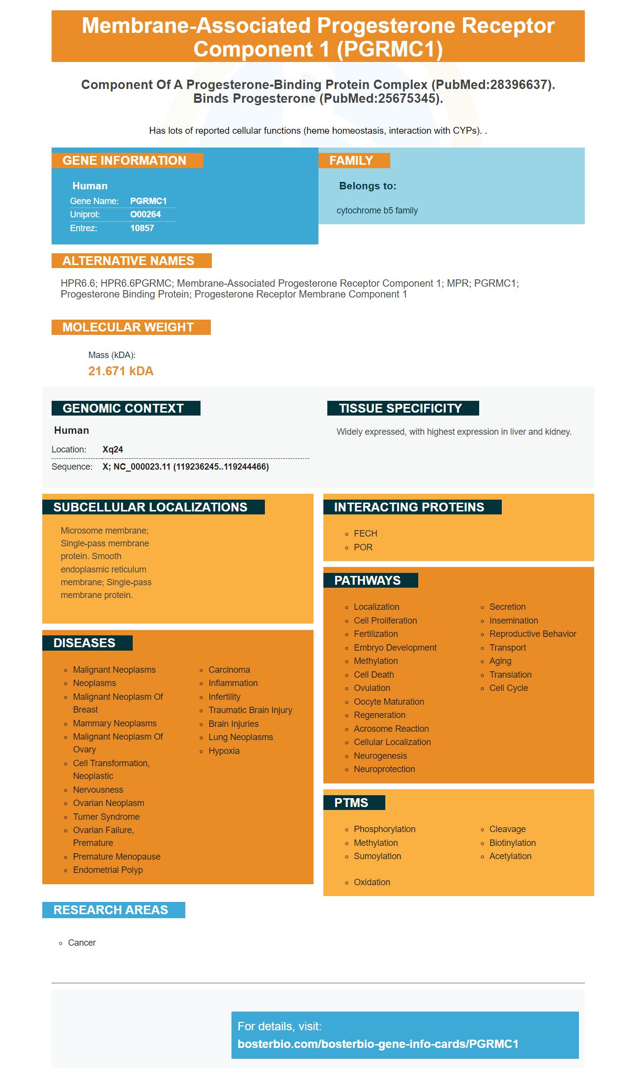
Facts about Membrane-associated progesterone receptor component 1.
Has lots of reported cellular functions (heme homeostasis, interaction with CYPs). .
Human | |
---|---|
Gene Name: | PGRMC1 |
Uniprot: | O00264 |
Entrez: | 10857 |
Belongs to: |
---|
cytochrome b5 family |
HPR6.6; HPR6.6PGRMC; membrane-associated progesterone receptor component 1; MPR; PGRMC1; Progesterone Binding Protein; progesterone receptor membrane component 1
Mass (kDA):
21.671 kDA
Human | |
---|---|
Location: | Xq24 |
Sequence: | X; NC_000023.11 (119236245..119244466) |
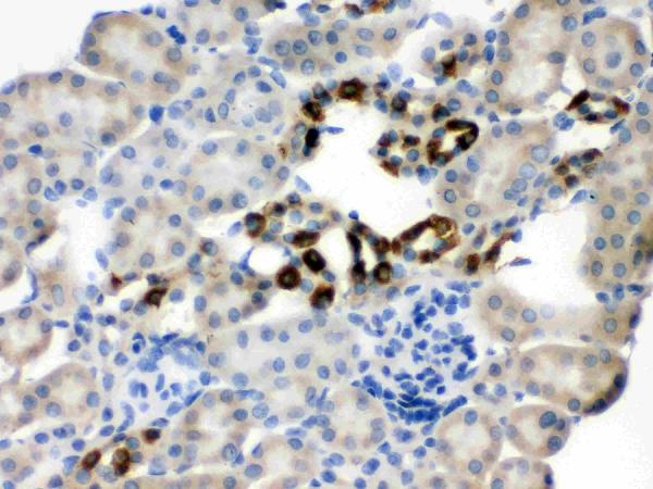
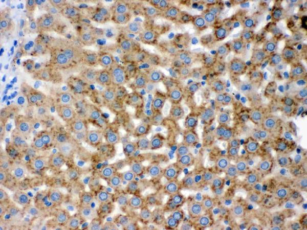
Widely expressed, with highest expression in liver and kidney.
Microsome membrane; Single-pass membrane protein. Smooth endoplasmic reticulum membrane; Single-pass membrane protein.
If you are a scientist looking for an inexpensive, yet effective way to detect and analyze gene expression, you should learn more about PGRMC1. This cargo-binding protein interacts with RTN3 and promotes degradation of a subset of MIDY proinsulin mutants. It also plays a role in cell morphology and behavior. We'll go over how to use this PGRMC1 marker.
The cargo-binding domain of Boster Bio PGRMC1 is critical for efficient ER phagosome clearance. While this domain must be located at the C-terminus of PGRMC1, other regions, such as the region downstream of residue 170, also contribute to substrate interaction. In this article, we discuss these findings. Also, we discuss how the PGRMC1 cargo-binding domain affects RTN3-dependent ER phagosome clearance.
In vitro, PGRMC1 is a type-II membrane protein with a long NTD and a short C-terminus. However, PGRMC1's long C-terminus should be cleaved by proteases while its short NTD remains intact. Therefore, when PGRMC1 is subjected to protease treatment, the cell blot will show a shift in mobility due to cleavage of the C-terminus. However, when PGRMC1-NTD antibody is used to detect PGRMC1, the signal of PGRMC1 is not affected. As a result, PGRMC1 is only at a negligible level on the surface of cells.
While PGRMC1 is essential for normal cell migration, phosphorylation may be detrimental to cell function. Mutations in the PGRMC1-HA protein are likely to lead to cell plasticity. In addition, the mutations in PGRMC1 cause cells to migrate with an irregular morphology and reduced ability to invade Geltrex pseudobasal membrane. Further, PGRMC1's phosphorylation state may alter cell morphology.
To determine if the MP cell line was effective in expressing the PGRMC1-HA protein, we first used a stable transfected MP cell line. Then, the MP cells were serum-starved for 2 h. Following this, a sterile p200 tip was used to scratch the monolayer, wash twice with PBS to remove floating cells, and add complete media. The images were acquired with a Nikon Eclipse Ti-U inverted phase microscope.
In this study, 3xFLAG-tagged POMC-6xHis was transiently expressed in HEK293T cells. After transfection, the membranes were analyzed by SDS-PAGE and immunoblotting using anti-FLAG antibodies. PGRMC1 siRNA treatment resulted in reduced HA expression. We are now able to distinguish between PGRMC1 and FLAG-tagged cargo proteins.
The PGRMC1 protein is a size-selective cargo receptor. Its activity as a receptor for misfolded substrates is a promising therapeutic target for ER-phagocytosis. Boster Bio's PGRMC1 marker was designed for these purposes. Its unique structure allows it to recognize cargos of the LMW type and has high affinity for the RTN3 protein.
The PGRMC1 gene is an important component of the ER, which is the cell's phagosomes. In addition, the gene controls the process of macroautophagy. It is involved in the degradation of proinsulin by the ER-phagy machinery. Hence, PGRMC1 has several applications. However, the PGRMC1 gene is not widely expressed.
The PGRMC1 protein recognizes species containing a proinsulin-A16P component of 150 kDa. This protein can be used as a phenotypic marker to determine the extent of mutant insulin in a tissue. However, in animal studies, PGRMC1 has been associated with various other diseases, including diabetes. For this reason, this gene is particularly useful for cell-based diagnostics.
The PGRMC1 protein is a selective phenotype that triggers the clearance of a subset of proinsulin mutants in the pancreas. It also plays a role in the degradative removal of MIDY proinsulin. Interestingly, the PGRMC1 protein is involved in a broader process, involving the ER protein BiP.
The boster protein capture was carried out using an anti-DDK affinity column and conventional chromatography steps. Using the boster protein capture method is applicable to all scientists worldwide. Further, the boster antibody will detect luminal cargo that is not normally degraded. This is the most promising approach for detecting luminal cargo in a wide variety of cell types. So, how do we use the PGRMC1 marker?
In this study, RTN3 is a bonafide ER transmembrane receptor and the ER transmembrane protein PGRMC1. It targets misfolded proinsulins. Moreover, it selectively targets smaller cargos compared to other reported ER-phagy substrates. In an experiment in mice, mutant proinsulins block the secretion of wildtype insulin, resulting in the development of insulin deficiency.
The PGRMC1 marker plays an important role in ER-phagy targeting. Mutations in this domain result in hyperphagia, a symptom of the diabetes syndrome MIDY. Other regions of the PGRMC1 c-peptide also participate in ER-phagy targeting, including residues downstream of 170.
The best uses of the PGRMC1 marker in a subset of MIDY pro insulin mutants are described. The PGRMC1 protein is associated with the degradative clearance of mutant proinsulin. The study demonstrates the importance of this marker in detecting ER stress and identifying the pathogens.
In this study, we used INS832/13 b-cells transfected with human WT or mutant proinsulins. After transfection, the b-cells were pulse-labeled with 35S-labeled amino acids for 30 min. Then, the cells were chased with complete media for 3 h. After the experiments, the cell lysate was subjected to a nonreducing Tris-tricine-urea-SDS-PAGE and phosphorimaging to detect oxidized proinsulin monomer.
The PGRMC1 marker targets C28F-POMC in the ER lumen, and loss of PGRMC1 reduces the sensitivity of the ER to endoH treatment. However, under the condition of knockdown of RTN3, C28F-POMC-FLAG still is sensitive to endoH treatment. Therefore, depleting PGRMC1 does not override the quality control of anterograde ER export.
The PGRMC1 marker was successfully used in 3 independent experiments with MIDY mutants. These mutants were evaluated in a WT or loss-A6/A11 proinsulin background. However, some of the MIDY mutants were unable to export from the ER in the wild-type background. Moreover, the PGRMC1 marker also prevented MIDY mutants from exhibiting a dominant-negative blockade of proinsulin export.
In addition to triggering degradation of a subset of MIDI-MIDY proinsulin mutants, the PGRMC1 marker has also been used as an effective biomarker. It recognizes species with proinsulin-A16P component of 150 kDa. It is a sensitive and specific biomarker for the characterization of insulin mutants.
In a recent study, researchers identified a new peptide marker, PGRMC1, which inhibits the degradative activity of a subset of MIDY-mutants. This marker is highly useful for identifying insulin-related genes and for predicting a patient's disease risk.
The PGRMC1-HA protein was expressed in MP cells. MP cells were seeded into a 24-well plate and serum starved for two hours before scratching the monolayer. After this, complete media was added. Images were captured using a Nikon Ti Eclipse confocal microscope. Cells were then counterstained with DAPI and photographed at 0 and 36 h.
The protein encoded by the PGRMC1 gene is phosphorylated by phosphorylating enzymes in the cytosol, which influences mitochondrial function. As a result, phosphorylating this protein may help identify a specific gene or RNA transcript. The protein is also expressed in a variety of cell types, including adipocytes, erythrocytes, and endothelial cells. The results of this study have important implications for the understanding of tumor development and cellular biology.
Although the phosphorylation of the PGRMC1 protein does not affect the properties of the actin cytoskeleton, phosphorylation of the protein has been associated with altered cell motility and morphology. These findings highlight the importance of understanding the molecular mechanisms involved in these processes and what they mean for the future of research. The PGRMC1 protein has multiple uses and can be a highly versatile marker for cell morphology and behavior.
The mitochondrial morphology and function of mitochondria are closely related. In addition to regulating mitochondrial function, PGRMC1 also influences cell morphology. In addition to the morphology of mitochondria, PGRMC1-HA expression is associated with an elongated or rounded cell form. The mitochondrial fragmentation pattern is associated with cardiovascular, neuromuscular, and aging processes.
The PGRMC1 gene is regulated by the estrogen receptor related 1 (ERR1) transcription factor, which is a steroid receptor. When ERR1 levels are suppressed, cell morphology and behavior change. Furthermore, the PGRMC1 phosphorylation state affects the abundance of proteins involved in protein folding. Ultimately, PGRMC1-induced changes in cell morphology can be used to identify the genetic basis of breast cancer.
In addition, the PGRMC1 gene is expressed in a wide range of cancer cells and is associated with poor patient outcomes. However, the PGRMC1 gene also interacts with the insulin receptor and glucose transporters, which are essential for the transition from aerobic to anaerobic metabolism in gestational diabetes. However, PGRMC1 is not the only gene that controls these processes.
PMID: 9705155 by Gerdes D., et al. Cloning and tissue expression of two putative steroid membrane receptors.
PMID: 11697142 by Bernauer S., et al. The human membrane progesterone receptor gene: genomic structure and promoter analysis.