This website uses cookies to ensure you get the best experience on our website.
- Table of Contents
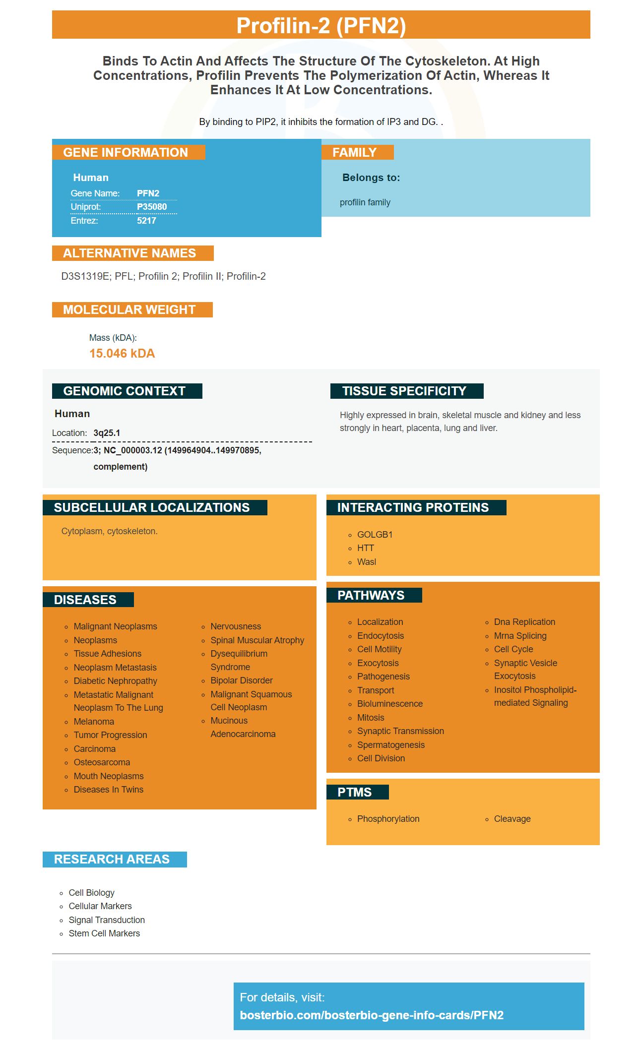
Facts about Profilin-2.

By binding to PIP2, it inhibits the formation of IP3 and DG. .
| Human | |
|---|---|
| Gene Name: | PFN2 |
| Uniprot: | P35080 |
| Entrez: | 5217 |

| Belongs to: |
|---|
| profilin family |

D3S1319E; PFL; profilin 2; Profilin II; profilin-2
Mass (kDA):
15.046 kDA

| Human | |
|---|---|
| Location: | 3q25.1 |
| Sequence: | 3; NC_000003.12 (149964904..149970895, complement) |
Highly expressed in brain, skeletal muscle and kidney and less strongly in heart, placenta, lung and liver.
Cytoplasm, cytoskeleton.


You have found the right place if you are looking for a biomarker that is reliable. Boster Bio has been producing high quality antibodies for over a decade. These antibodies have been tested against positive and negative samples to ensure their high specificity. Boster offers product credits to early reviewers of new products and rewards scientists around the world with product credits. Here are some of the benefits of boster Bio:
In studies of lung carcinoma, a monoclonal cell expressing the PFN2a protein is used. The C2C12 cell lines resulting have been transformed using CRISPR/Cas9 to express PFN2a genes. Because it suppresses histone deacetylase activity, the PFN2a genes can be used for research in lung cancer.
PFN2a's role is still unclear. The protein is involved in myogenic development and regulates p53 through its inhibition of acetylation. However, it has been shown to inhibit p53 and the p53 pathway, and it may also contribute to the regulation of muscle development. This gene could play a role the regulation of C2C12 myoblasts.
The gene encodes the protein MyHC. The PFN2 gene is expressed by a number of different cell types, including stem cells and embryonic fibroblasts. In some studies, the level of myogenin (or myHC) in a cell is related to the expression of PFN2. In C2C12 cells, PFN2a also decreases the number MyHC-positive and myogenin positive cells.
In C2C12 cells, overexpression of PFN2a promotes caspase 3 activity and caspase 8. In addition, the overexpression of PFN2a inhibits C2C12 cell proliferation and increases p53 expression. In C2C12 cells, the PFN2aGFPGFP mRNA was detected to increase in myoblasts. This protein also inhibits sarcomere assembly and myogenic differentiation, suggesting it has a beneficial effect in tumorigenesis.
The mRNA PFN2a, which is present in the cell cytoplasm on differentiation day 3, promotes apoptosis. It also inhibits HDAC1 activity and suppresses myogenic differentiation in C2C12 cells. It has been shown to promote the expression of p53 in C2C12 C2C12 Myogenic Stem Cells. PFN2a promotes the expression p53 in glioma-cells.
As PFN2a is essential for myogenic differentiation, it is essential to identify the regulator of apoptosis. Molecular markers of apoptosis are essential for myogenic differentiation, so it is important to identify this regulator to determine the optimum cell number. To test this hypothesis, we found that PFN2a cells with the gene overexpression decreased proliferation in C2C12-derived cells. It was important that both PFN2a cells were grown at the same density on differentiation day 0, and analyzed for late-apoptosis.
HDAC1 is an important regulator for the G1–to–S phase transition. This is vital for normal cell cycle progression. Both HDAC1 (and HDAC2) play an important role in cell division. This enzyme inhibits p57, p21. HDAC1 plays a key role in cell proliferation regulation. HDAC1 as well as HDAC2 can be found in cancer cells.
Although HDAC1 expression in tumor cells has been studied in bulk tissues, previous studies only focused on cell lines from one patient and did not consider the function of individual HDACs. This study, however aimed to identify the individual HDACs of GBM GSCs. The results eventually led to isoform selective HDACi. Here, we review the key data relating to HDAC1 in cancer stem cells.
A00256-2 was the name of the corresponding antibody. It is designed to recognize HDAC1 close to the C-terminus in human DNA. The anti-HDAC1 antibody reacts with Human, Mouse, Rat, and Mouse. It is suitable for immunofluorescence experiments and western blotting. It can recognize HDAC1 in all three cell lines when used in WB applications. It can also be used in a variety of other applications.
HDAC1 protein is required for p57 or p21 upregulation. These proteins cannot regulate the cell's cycle without HDAC1 or HDAC2. The cells will cease to multiply and eventually undergo apoptosis. HDAC1 is also vital for the proper functioning p57/p21. Its absence can cause abnormal cell cycle profiles. The HDAC inhibitor LSD690 can stop the activity of SE in cell line cells at levels that cause cell death.
HDAC1 (and HDAC2) have distinct targets. Further experiments will help to determine their target preferences. However, this is an illustrative example of a protein that is essential for normal cell development. Boster Bio HDAC KO cells require both HDAC1 & HDAC2 to ensure proper cell differentiation. These enzymes can cause significant cell changes that will disrupt growth and development.
The PFN2 markers encode a profilin-like protein. This gene is located on chromosome 3q25.1 and is believed to regulate actin polymerization. The PFN2 genome is available in two forms. One encoding a human protein while the other a E.coli-derived protein. The PFN2 protein interacts with the cytoskeleton to modulate cell division and movement. PFN2 inhibits the activity of DG and IP3, two proteins that affect the cytoskeleton.
The PFN2 subset of NS5A genes has been implicated with a wide range of diseases. Chinese doctors have discovered the PFN2 genome is essential for the development of cancer. Although the PFN2 genes is the most prevalent cause of cancer in humans today, researchers are still not certain of its exact role in cancer. For confirmation, a test is required to detect the PFN2 genes.
The PFN2 marker, which is an important transcription factor, regulates cell growth, differentiation, and apoptosis. PFN2a plays a key regulatory role in cell growth, apoptosis, and differentiation. It is important to keep in mind that the protein only expresses in cells in the s-phase. This phase mainly conducts DNA replication and prepares cells for cell division. A transcription factor, PFN2a also inhibits C2C12 Myogenic Cell Growth.
Boster bio anti PFN2 antibody was made to detect PFN2 levels in cells, tissue, blood, and other samples. It reacts with Human, Mouse, Rat, and ICC proteins. The antibody is highly specific and exhibits high affinity. Boster Bio's anti-PFN2 product has been validated for use in immunohistochemistry, Western blotting, and ELISA.
Overexpression of the PFN2a gene promotes the production and activation of caspase 8, caspase 3, in C2C12 myogenic cellular cells. This results in a reduction of viable cells and an increase in early apoptosis. PFN2a is also known to inhibit myogenic differentiation and increase p53 expression in cells. PFN2a, although essential for maintaining healthy tissue and other functions, is not well-known.
PFN2a is a potent inhibitor of HDAC1 and promotes cell death. It is associated to myogenic development and has an affect on HDAC1 concentrations in C2C12 cellular cells. The PFN2a genes regulate the activity p53. The PFN2a protein is required for normal cell growth and differentiation, so it is important to analyze the gene's expression in cells that express the gene.
Research has shown that PFN2a is involved in the regulation of the assembly and maturation of mature sarcomeres. Overexpressions of PFN2a were found in C2C12 cells to cause disordered MyHC/aActinin protein expression. Furthermore, PFN2a downregulates tropomyosin 1 and myogenic differentiation in C2C12 cells.
The PFN2 marker has a wide range of uses, from the identification of a cellular cytotoxic agent to the development of therapeutic antibodies. In cancer research, it is important to understand the mechanism of cell death. PFN2 markers can be used to detect early signs of cancer. You should consider this new diagnostic tool for PFN2 research.
PMID: 8365484 by Honore B., et al. Cloning and expression of a novel human profilin variant, profilin II.
PMID: 11027290 by Lambrechts A., et al. Profilin II is alternatively spliced, resulting in profilin isoforms that are differentially expressed and have distinct biochemical properties.