This website uses cookies to ensure you get the best experience on our website.
- Table of Contents
1 Citations 6 Q&As
2 Citations 6 Q&As
40 Citations 16 Q&As
41 Citations 16 Q&As
17 Citations 15 Q&As
11 Citations 15 Q&As
10 Citations 15 Q&As
20 Citations 16 Q&As
8 Citations 3 Q&As
8 Citations 3 Q&As
9 Citations 5 Q&As
31 Citations
1 Citations
Facts about Platelet endothelial cell adhesion molecule.
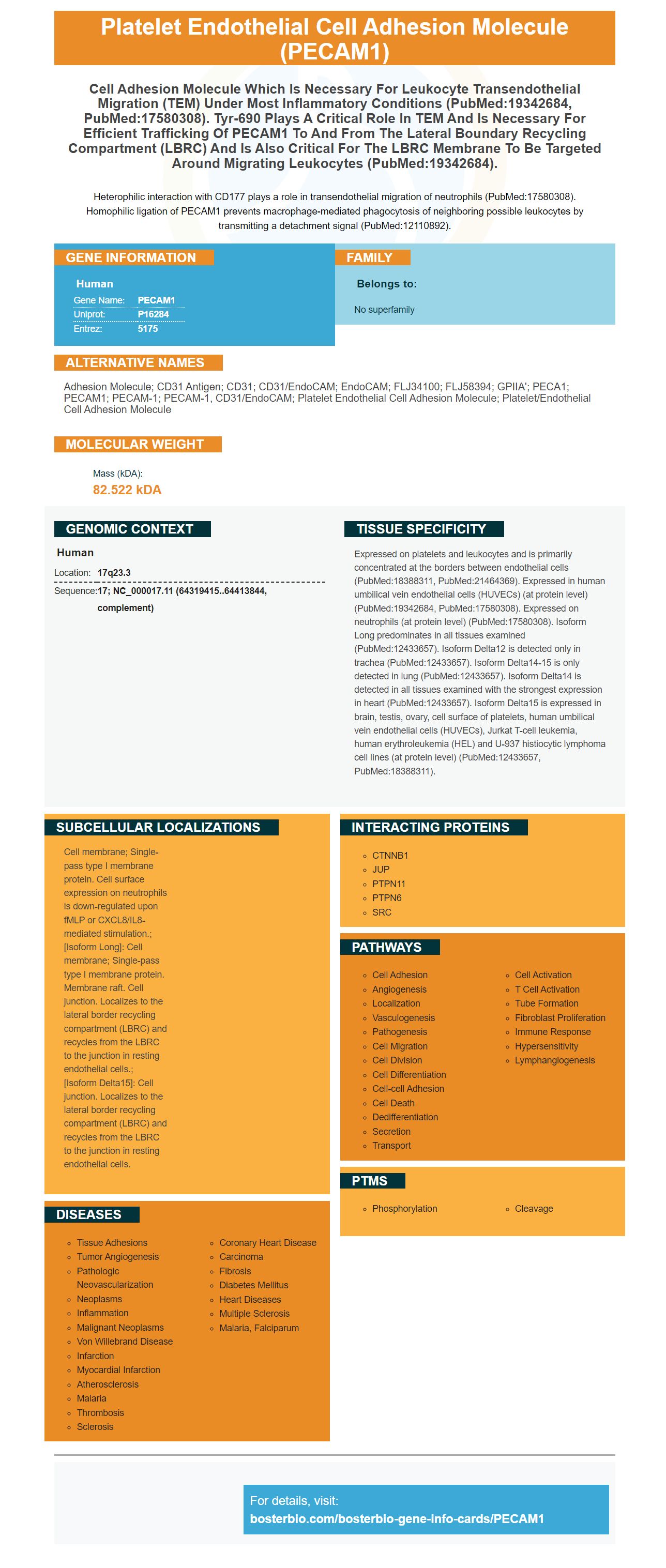
Heterophilic interaction with CD177 plays a role in transendothelial migration of neutrophils (PubMed:17580308). Homophilic ligation of PECAM1 prevents macrophage-mediated phagocytosis of neighboring possible leukocytes by transmitting a detachment signal (PubMed:12110892).
Human | |
---|---|
Gene Name: | PECAM1 |
Uniprot: | P16284 |
Entrez: | 5175 |
Belongs to: |
---|
No superfamily |
adhesion molecule; CD31 antigen; CD31; CD31/EndoCAM; EndoCAM; FLJ34100; FLJ58394; GPIIA'; PECA1; PECAM1; PECAM-1; PECAM-1, CD31/EndoCAM; platelet endothelial cell adhesion molecule; platelet/endothelial cell adhesion molecule
Mass (kDA):
82.522 kDA
Human | |
---|---|
Location: | 17q23.3 |
Sequence: | 17; NC_000017.11 (64319415..64413844, complement) |
Expressed on platelets and leukocytes and is primarily concentrated at the borders between endothelial cells (PubMed:18388311, PubMed:21464369). Expressed in human umbilical vein endothelial cells (HUVECs) (at protein level) (PubMed:19342684, PubMed:17580308). Expressed on neutrophils (at protein level) (PubMed:17580308). Isoform Long predominates in all tissues examined (PubMed:12433657). Isoform Delta12 is detected only in trachea (PubMed:12433657). Isoform Delta14-15 is only detected in lung (PubMed:12433657). Isoform Delta14 is detected in all tissues examined with the strongest expression in heart (PubMed:12433657). Isoform Delta15 is expressed in brain, testis, ovary, cell surface of platelets, human umbilical vein endothelial cells (HUVECs), Jurkat T-cell leukemia, human erythroleukemia (HEL) and U-937 histiocytic lymphoma cell lines (at protein level) (PubMed:12433657, PubMed:18388311).
Cell membrane; Single-pass type I membrane protein. Cell surface expression on neutrophils is down-regulated upon fMLP or CXCL8/IL8-mediated stimulation.; [Isoform Long]: Cell membrane; Single-pass type I membrane protein. Membrane raft. Cell junction. Localizes to the lateral border recycling compartment (LBRC) and recycles from the LBRC to the junction in resting endothelial cells.; [Isoform Delta15]: Cell junction. Localizes to the lateral border recycling compartment (LBRC) and recycles from the LBRC to the junction in resting endothelial cells.
If you're looking for a reagent to measure the presence of the PECAM1 protein, you have come to the right place. This article will cover the biological activity as well as the validation and advantages of the PECAM1 marker. The medical field has many applications for the PECAM1 gene marker, as you'll discover. Here are some examples. Find out more! Don't forget to share your research findings and ideas with other scientists!
The Boster Bio Anti CD31/PECAM1 monoclonal antibodies have a variety of applications. They interact with Human, Mouse, and Rat CD31 and share 68 percent amino acid sequence homology. The Boster Bio Anti CD31/PECAM1 Marker is made from whole rabbit serum. It can be utilized in a variety of cell-based applications and has a wide variety of applications.
PECAM-1 is an extracellular molecule expressed by platelets, endothelial cells and a few lymphocyte subsets. It connects to the surface of vascular cells and helps to promote their adhesion and function. In addition to adhesion PECAM-1 might also interact with other molecules, such as integrin alpha 5 beta 3.
Studies have revealed that PECAM-1 plays a significant role in the development of endothelial cell barriers. It blocks circulating leukocytes and platelets from adhering to the endothelial cell junctions and helps stop platelets from adhering to them. PECAM-1 also inhibits Bax-induced cells death. It has been demonstrated to be involved in thrombosis and inflammation, as well as cancer.
PECAM-1 is a molecule that signals and adhesion molecule that is used by blood vessels. It is vital in maintaining and rebuilding the vascularpermeability barrier. It is also a stress-response molecule that preserves the integrity of junctions between endothelial cells and facilitates the permeability of the vascular system. It is a well-known treatment for disorders of blood-brain barrier.
As a critical component of the endothelial barriers, PECAM1 could provide a new therapy option for the treatment of sepsis and ARDS. In one study, mice that had deficient PECAM1 were transplanted with bone marrow cells express PECAM1, which led to improved resistance to endotoxic shock and maintained the integrity of the vascular permeability barrier.
The complex PECAM-1 and SHP-2 is an important component in the TEM. It is crucial for efficient trafficking PECAM1. It also prevents phagocytes from ingestion of tightly apposed, viable cells. This protein plays an essential role in the development and progression of endothelial disease such as tumor angiogenesis and hemangiomas, and hemangiomas. When it is present in high quantities in blood, it may be targeted to the cell and control its growth and movement.
Intercellular junctions control the cellular expression of PECAM-1. Intercellular junctions regulate leukocytes' trafficking, mechanotransduction and cell proliferation. It regulates vascular permeability and also maintains the integrity of the vascular system through resistance to mechanical forces under shear stress. It may also play a role as mediator of thrombosis, inflammation and immune responses. Further research is needed to better understand the physiological roles of this marker.
The first recognition of PECAM-1 was made within the context of the search for new adhesion molecules in ECs. It was later discovered to be distributed on the cell's surface, preferentially near cell-cell contacts. However, further studies showed that there are two isoforms of PECAM-1 with distinct functional roles. Yan et al. studied the functional consequences of muPECAM-1 cDNAs with alternative muPECAM-1 by transfecting L cells with the cDNA of either. The authors found that PECAM-1 isoforms interact differently to ECs and have different effects on their functions.
The PECAM-1's cytoplasmic region is composed of eight separate exons. The exon 14 regulates PECAM-1's interaction with intracellular proteins and also modulates its extracellular adhesive properties. Transfected cells that contain FL muPECAM-1 and exon 14 exhibit heterophilic aggregation dependent on calcium. The PECAM-1 isoforms with exon 14 play a role in facilitating calcium-dependent homophilic aggregation. The ligand specificity of PECAM-1 is affected by the absence of Tyr-686 from exon 14.
Studies on PECAM-1 have revealed that it is an essential element of the barrier between endothelial cells and vascular cells. The research has shown that antibody-PECAM-1-binding IgD6 can enhance the homophilic affinity of this receptor, which may have translational benefits. A plethora of studies has confirmed that mice lacking PECAM-1 do not exhibit vascular abnormalities when they are resting however they exhibit a severe appearance when exposed to bacteria.
The PECAM-1 molecule serves a variety of functions, including the regulation of inflammation and signal transduction. PECAM-1 is an endothelial cells-biology protein. It also functions as a stress-response protein protecting the integrity of the junction and improving the permeability of blood vessels. These functions are further described in this chapter. PECAM-1 is a crucial chemical that promotes the growth of endothelial cells, but it is also vital for the transendothelial flow of leukocytes.
The PECAM1 antibody serves dual purposes. It reduces the production of cytokine, and also maintains the integrity of vascular walls. To fully comprehend the mechanism behind PECAM1 anti-body activity, further research is required. Once it has been validated, it is then able to be used for the diagnosis and treatment of chronic inflammatory conditions. In this article, we will describe the process of validation for the PECAM1 antibody. We also discuss the possible applications for this immunotherapy.
To confirm the PECAM1 marker We first selected genes that are highly expressed in hepatitis. PECAM1 expression is crucial for cell apoptosis as well as the inflammation response. The same function is played by this marker in the dental pulp tissues of rats and HDPFs. We were able validate the target cells with this marker. Our results show that PECAM1 is extremely expressed in patients suffering from hepatitis C.
We also found that patients who suffered from sepsis-induced respiratory injury had higher levels of sPECAM1 and pulmonary damage. These findings suggest that sPECAM1 could be a useful biomarker for ARDS or a marker for early VILI risk. Further research is needed to confirm that PECAM1 is actually a reliable marker.
In addition, we showed that human effector and memory CD4+ T cells do not require PECAM-1 to function in the BBB. This result was confirmed with staining for VE JAM, occludin and JAM. The junctional structure of PECAM-1/ pMBMECs was also unaltered. They also have impaired barrier properties. Additionally their barrier properties did not alter their molecular structure in general.
It is interesting to note that the PECAM1 marker and CXCR4 have been shown to interact. The two markers interact with each other, indicating their co-expression. The study also found that CXCR4 and PECAM1 are bound to CXCR4 within HDPF tissues. Additionally this marker interacts with CXCR4 and MEF2C and MEF2C. These are two related genes that are part of the immune system.
Researchers compared the PECAM-1 protein expression levels in lesions from active and initial MS patients to normal white tissue of healthy people in this study. They also evaluated the expression of the PECAM-1 gene to cortical gray matter from patients with no MS. These preliminary findings suggest that PECAM-1 might play an important role in MS. The results of this study are in line with the findings of a previous method.
The PECAM1 marker is an effective treatment for cancer. The marker is present in the endothelium vascular. It has been linked in the progression of metastatic cancers. It is believed to function by altering the tumor's microenvironment and stimulating the proliferation of tumor cells. However, its effect has not been confirmed in the context of tumor angiogenesis. The goal of this study was to determine if functional PECAM1 promotes the proliferative phenotype of the melanoma cell line by co-culture with mouse endothelium.
The PECAM-1 marker is used to detect metastatic tumors. Studies have revealed that PECAM-1 could influence the activity of TIMP-1. Additionally, it can be used as a marker for prognosis. PECAM-1 is known to regulate expression of TIMP-1 in metastatic cancers. More research is required to prove its effectiveness in treating cancer. It could be useful in the treatment of metastatic cancers.
The PECAM1 marker has many benefits that have been established in animal studies. The protein has a positive role in preventing sepsis as well as ARDS by inhibiting the growth of inflammatory cells, and also restoring endothelial continuity. Recently, a study by Maas et al. found that mice lacking PECAM1 bone marrow cell lines showed decreased rates of endothelial cell-to-endothelial permeability barrier reversal.
The PECAM1 marker is an excellent prognostic tool to detect skin tumors. It has been demonstrated to differentiate benign tumors from malignant. PECAM-1 is a biomarker that can be used to assess the dangers of Melanoma. However, its use could cause a problem with the diagnosis of tumors in the soft tissues deep. Histiocytes exhibit weak staining of the protein.
Although PECAM-1 is an excellent biomarker for early pregnancy, it is not an accurate predictor of fetal loss for all women. Although there have been few reports on the relationship between PECAM-1 and pregnancy loss However, the results of previous studies have revealed that the coexistence of these two polymorphisms increase the risk of miscarriage. Moreover, the PECAM-1-373G allele is associated with a greater risk of miscarriage in younger women.
PMID: 2351935 by Simmons D.L., et al. Molecular cloning of CD31, a putative intercellular adhesion molecule closely related to carcinoembryonic antigen.
PMID: 1700999 by Stockinger H., et al. Molecular characterization and functional analysis of the leukocyte surface protein CD31.
*Showing only the more recent 20. More publications can be found for each product on its corresponding product page