This website uses cookies to ensure you get the best experience on our website.
- Table of Contents
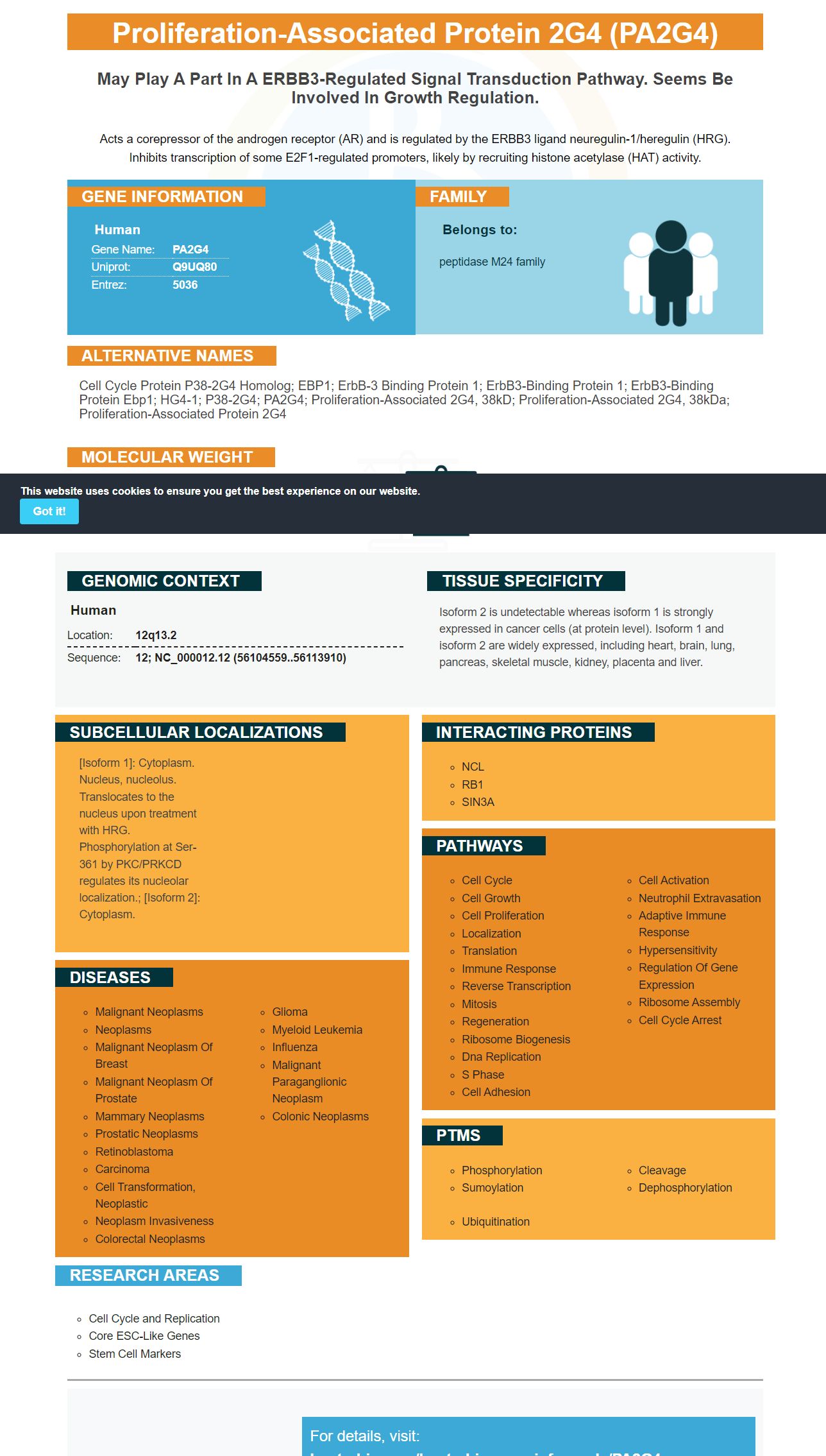
Facts about Proliferation-associated protein 2G4.
Acts a corepressor of the androgen receptor (AR) and is regulated by the ERBB3 ligand neuregulin-1/heregulin (HRG). Inhibits transcription of some E2F1-regulated promoters, likely by recruiting histone acetylase (HAT) activity.
Human | |
---|---|
Gene Name: | PA2G4 |
Uniprot: | Q9UQ80 |
Entrez: | 5036 |
Belongs to: |
---|
peptidase M24 family |
Cell cycle protein p38-2G4 homolog; EBP1; ErbB-3 binding protein 1; ErbB3-binding protein 1; ErbB3-binding protein Ebp1; HG4-1; p38-2G4; PA2G4; proliferation-associated 2G4, 38kD; proliferation-associated 2G4, 38kDa; proliferation-associated protein 2G4
Mass (kDA):
43.787 kDA
Human | |
---|---|
Location: | 12q13.2 |
Sequence: | 12; NC_000012.12 (56104559..56113910) |
Isoform 2 is undetectable whereas isoform 1 is strongly expressed in cancer cells (at protein level). Isoform 1 and isoform 2 are widely expressed, including heart, brain, lung, pancreas, skeletal muscle, kidney, placenta and liver.
[Isoform 1]: Cytoplasm. Nucleus, nucleolus. Translocates to the nucleus upon treatment with HRG. Phosphorylation at Ser-361 by PKC/PRKCD regulates its nucleolar localization.; [Isoform 2]: Cytoplasm.
The PA2G4 marker is a high-affinity primary antibody available from Boster Bio. Its unique sequence domain is 1-394aa. The PA2G4 protein is expressed in E.coli and has a His-Tag. The protein is stable for up to a week at room temperature, but should be stored at -20degC to -80degC for long-term storage. However, it should be stored at -20degC to -80degC to avoid repeated freeze-thaw cycles. Scientists can earn product credits by sharing their results, which can earn them rewards. All scientists can participate.
Boster Bio offers high-affinity primary antisera against the PA2G4 marker, a chromosomal marker for the human cell membrane. These antibodies have been developed by Steven Boster, a former graduate student who earned the nickname "he who converts science in the lavatory" because of his tenacity and commitment to achieving high-quality and precise results. In fact, Boster has developed proprietary ELISA platforms, such as PicoKine(tm) - to deliver high-sensitivity ELISA kits.
The new Membrane Staining kit from Boster Bio can help researchers maximize the efficiency of protein transfer. This kit uses an enzyme-conjugated secondary antibody to detect protein bands on nitrocellulose or PVDF membranes. Ponceau S is a negative-charged dye that binds to amino-acid residues to produce protein signals. The staining process is rapid and easy, and it is suitable for detecting protein bands on both nitrocellulose and PVDF membranes.
To determine the protein transfer efficiency, the membrane was stained with a lectin that is specific to the ligand. AAL, PHA-E, and SNA lectins were used to identify protein bands. After incubation, the gel was washed with TBST three times, and the lectins were visualized using ECL Plus reagents (Beyotime). The membrane was stained by immersion in a 50/50 methanol/water mixture. The exposure time for the membrane was the same as for the traditional fixation method.
In addition to immunoglobulin G, the lectin is used to detect proteins in plasma. The method used for plasma and blood serum separation is also highly efficient. It enables researchers to detect two targets in one run, saving both time and money. Furthermore, the membrane staining procedure can be used to test the effectiveness of the antibody re-probed against multiple targets simultaneously.
Nitrocellulose Membrane is one of the most common protein-blotting matrix options available. Its high protein-binding affinity makes it compatible with various detection methods. It also immobilizes proteins, glycoproteins, and nucleic acids. Its high-binding capacity allows it to be widely used in direct dot binding assays and electroblotting. If you're looking for the most effective way to immobilize proteins on electroblot membranes, the nitrocellulose membrane from Boster Bio can help.
A recent study has demonstrated that chicken antibodies tend to bind nylon-based membranes. Switching to nitrocellulose membranes reduces background staining. Furthermore, Ponceau Red stock, made from 2% Ponceau S in 30% trichloracetic acid, improves the efficiency of protein transfer by membrane staining. However, the Ponceau Red stock requires a different procedure.
In addition to the sensitivity of the gel and the type of membrane used, the efficiency of protein transfer can also depend on the molecular weight of the transferred protein. The higher the molecular weight, the higher the hydrophobicity of the protein molecules. This will affect the type of membranes suitable for detecting small molecular weight proteins. In addition, the higher the molecular weight, the greater the sensitivity of the WB method.
The detection sensitivity of the western blotting process is affected by the efficiency of protein transfer and the loss of proteins. The loss of proteins may lead to inconsistent results, and the use of detergents that remove hydrophilic proteins can affect the reproducibility of the analysis. The new Boster Bio solution improves protein transfer efficiency by membrane staining
The PA2G4 marker is a novel genetically encoded protein that has many uses. This protein contains multiple phosphorylation sites and a number of lysine-rich regions. Mutagenesis studies have helped to determine its functions. The structure of the PA2G4 protein reveals three lysine-rich regions, one near the N terminus, another near loop 1 and a third at the C terminus. Mutation studies have also helped to define the role of the PA2G4 protein in nucleocellular localization.
The PA2G4 protein interacts with p53 E3 ligase HDM2. This interaction promotes p53 polyubiquitination and degradation, both of which lead to reduced p53 activity. According to Kim et al., the PA2G4 protein stabilizes HDM2, which in turn facilitates its interaction with Akt. Thus, PA2G4 is useful in cancer research.
The PA2G4 protein is a unique genetic marker because it lacks hydrolyzing activity on MAP substrate. As a result, PA2G4 adopts a hMAP2-like structure, which is essential for ribosome biosynthesis. Moreover, the PA2G4 gene is highly conserved across species. The gene coding for PA2G4 is located in the central nervous system of human cells, which makes it useful for identifying genetic disorders.
The expression of PA2G4 correlates with a patient's prognosis in human cancer. In cancer, PA2G4 is expressed by cells in response to ERBB3 signaling. The two isoforms of the protein have different functions and can have opposing roles depending on their context. For instance, the ERBB3-binding protein 1 (ERBB3) in tumor cells has an oncogene-like role in cancer cell proliferation.
PA2G4-p42 isoforms are unable to undergo polyubiquitination, and the E3 ubiquitin-protein ligase Bre1 binds phosphorylated Ser360 in PA2G4-p48. Therefore, PA2G4-p48 and FBXW7 share similar roles, although their interactions are somewhat different. Interestingly, the PA2G4-p42 isoform isoforms still contact FBXW7.
In addition to PA2G4 and ERBB2, EFTUD2, GAPDH and NOP56 showed significant associations with poor overall survival in melanoma patients. The expression of PA2G4 showed a marked up trend with increasing pathological or T stages. These gene expression levels were analyzed using Kaplan-Meier survival curves, with a median cutoff point that differentiated between the low and high-expressing groups.
PMID: 9345902 by Lamartine J., et al. Molecular cloning and mapping of a human cDNA (PA2G4) that encodes a protein highly homologous to the mouse cell cycle protein p38-2G4.
PMID: 10682683 by Yoo J.Y., et al. Interaction of the PA2G4 (EBP1) protein with ErbB-3 and regulation of this binding by heregulin.