This website uses cookies to ensure you get the best experience on our website.
- Table of Contents
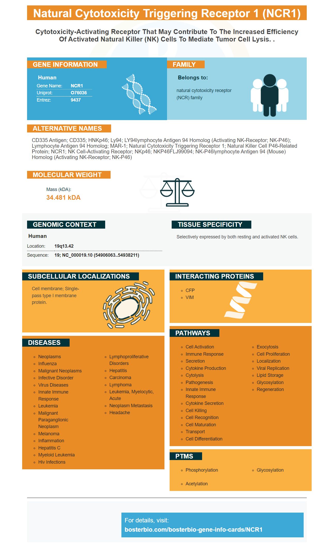
Facts about Natural cytotoxicity triggering receptor 1.
Human | |
---|---|
Gene Name: | NCR1 |
Uniprot: | O76036 |
Entrez: | 9437 |
Belongs to: |
---|
natural cytotoxicity receptor (NCR) family |
CD335 antigen; CD335; hNKp46; Ly94; LY94lymphocyte antigen 94 homolog (activating NK-receptor; NK-p46); Lymphocyte antigen 94 homolog; MAR-1; natural cytotoxicity triggering receptor 1; Natural killer cell p46-related protein; NCR1; NK cell-activating receptor; NKp46; NKP46FLJ99094; NK-p46lymphocyte antigen 94 (mouse) homolog (activating NK-receptor; NK-p46)
Mass (kDA):
34.481 kDA
Human | |
---|---|
Location: | 19q13.42 |
Sequence: | 19; NC_000019.10 (54906063..54938211) |
Selectively expressed by both resting and activated NK cells.
Cell membrane; Single-pass type I membrane protein.
The NCR1 marker is a key component in the NK cell's arsenal of cytotoxic substances. This marker also exhibits expression on lymphocyte subsets different from NK cells. This article explains how T-cells which express N1/28z are able to target cancerous cells. This marker is worth learning more about. Boster Bio: The Best uses of the NCR1 marker
The NCR1 receptor is found on the surface of a variety of lymphocytes and is previously thought to be a particular NK cell marker. The receptor was found to be expressed on subsets of lymphocytes which are not NK cells. Researchers have found that NCR1 is present in lymphocytes that are malignant, such as those from patients suffering from Sezary syndrome. The receptor is identified to play an inhibitory role. It is possible that the NCR1 receptor may be involved in the growth of leukemia or NKT cells.
It has been proven that cancer cells increase the expression of NCR1 which allows them to recognize NK cells. However, the precise cell ligands of NCR1 remain unknown. The in vivo test has provided preliminary insights into its therapeutic potential. The researchers also discovered that the NCR1 receptor was associated with an increase in the inflammatory response.
The NCR1 receptor is highly expressed on the surface of human T cells and has the ability to target a variety of cancers. The non-MHC restricted nature of the receptor could facilitate strategies that target different malignancies with a single antibody. As a result, it is regarded as an "universal" treatment for immunotherapy against cancer. Furthermore, NCR1 expression has been found to be decreased in cancer patients and inhibited by inhibitor ligands expressing in tumors. These inhibitors may be bypassed in the event that NCR1-transduced T cells are employed instead.
Boster Bio may use NCR1 receptor to recognize NK cells. The receptor could be associated with the endocytosis process as well as the recycling of extracellular matter. However, the researchers discovered that a number of cell-like spherical body structures in WML and Parkinson's disease are associated with the presence of NCR1 receptor. Fluorescent microscopy further confirmed the structure.
In this study, a subset of CD56bright/CD16lo/-/NKp46+ cells was discovered. These cells constitute between five to ten percent of total population. The CD56brightCD16lo/cell subset of CD56bright/CD16+NK cells is composed of CD56dim//NKp46+ cells. Additionally, the CD56dim/CD16lo/ cells were significantly lower in the proportion of CD56+/CD5+ T cells.
The combination of genetically modified T-cells as well as the NCR1 receptor as a NK cell marker is an effective way to enhance the capacity of NK cells to recognize cancer cells. These studies will be used to confirm the use of NCR1 as a NK-cell marker in more extensive studies. It's now time to use NCR1 receptor from Boster Bio as an marker for NK-cells.
The immune system has many functions for the NK cell. As part of the immune system's innate system, the NK cells are responsible for maintaining the health of tissue stromal cells. They aid in regulating CNS immunity. This means that their presence in the body can aid in preventing the development of autoimmune diseases. It is essential to identify the NK receptors within these tissues, since the immune system is the key to fighting infections.
Numerous studies have demonstrated that CD8+ NK cells are associated with clinical indicators in chronic HIV-1 infection. These findings have important clinical implications. These cells could be a novel NK-cell derived innate immune correlate. The expression of CD8+ NK cells in peripheral blood may be useful in predicting outcomes in clinical care. This article explains this phenomenon in depth. Also, we discuss the role played by the CD8+ NK cells in HIV infection.
The genes NKp30 and NKG2A are expressed in NK cells, however their frequencies differ among the subsets. NK cells that express NKp30 and NKG2A have higher frequency of these antigens that those that express both receptors. CD8-NK cells express both receptors with high frequency and the findings of this study will allow us to identify these subsets.
The CD8+ subset possesses more functional properties than CD8 cells. These cells are able identify and kill tumor cells. They also aid in stopping the spread of infections. CD8+ cells are more vulnerable to cytotoxic effects , but they also possess a greater proliferative capacity. They also express killer Ig-like receptors. They are transmembrane proteins that play important functions in the immune system.
Moreover it has been observed that both aNKR and CD3 antigen are expressed in varying amounts on human lymphocyte subsets. These findings suggest that distinct Granzymes play different roles in regulating and responding to immune responses. However, they also highlight the importance of understanding the mechanisms that drive the expression of CD3 in human cells. To better know the mechanism by which CD3 is expressed in lymphocyte subsets it is first necessary to understand what CD3 antigen is doing for them.
We utilized immunostaining and immunostaining to detect CD3 and CD8 in the NK cells. We then used a specific cytokine for assessing the expression of CD8+ cells by using staining with antibodies. We also used degranulation staining for evaluating intracellular cytokine levels in different lymphocyte subsets. This was done to determine whether these cells were active in the NK immune response.
A similar analysis was carried out to determine which CD8+ NK cells are more sensitive to the four functions NK cells perform. The highest levels of expression were observed in NKT cells, while the smallest levels were found in NK cells. Additionally, the levels of FSC were higher in NK cells, compared to CD3+CD56+ T cells. The same increase in CD8+ NK cells was seen in untreated subjects.
Functional NK cell assays were performed using the same methodology as described in previous studies. The experiments included stimulation of 105 sorted NK cell lines with 10 ng/ml IL-15 , and K562 cells in a 2:1 ratio. The cells were fixed and stained with anti-IFN-g Pacific Blue. Antibodies to CD8 and CD86 were also used to detect intracellular expression of cytokines.
We discovered that the anti-tumor activity of T-cells expressing N1/28Z was enhanced in human primary T-cells that have an atypical antigen receptor. These results suggest the possibility of using the same non-MHC restricted receptor to target multiple malignancies. The chimeric receptor blocks the tumor inhibitor ligands that could allow strategies to target multiple malignancies at once.
Transduced lymphocytes that express N1/28z and NGFR were co-cultured in same environment as CFSE-labeled cancer cells. They were then gated to a particular tumor cell population by flow cytometry after 18 hours. After the tumor had growing, a proportion of CFSE labeled CFSE TB-cells were identified and analyzed with the Winn assay.
Haanen and his colleagues studied a previously developed CAR T-cell product in phase I and II clinical trials. This product targets CLDN6, which is a tumor-specific antigen that is that is expressed in a variety of solid tumors, but is silent in normal adult tissue. Both treatments increased the growth of CAR T cells transferred and decreased the death rate of tumor cells. This combination could help improve the outcomes of hematologic malignancies.
These cells showed enhanced anti-tumor efficacy , which led to an increase in IL-17 secretion. The CAR-T cells would not survive in the presence Tregs as they'd be lacking IL-2 to sustain their activity. However, the same strategy can be employed in conjunction with traditional co-stimulatory endodomains. They could be engineered to express any anti-tumor molecule.
Another approach that is promising for the use of CAR-T cells is to engineer of macrophages to express CAR T cells that target TAMs. CAR-T cells could be able to re-educate cancer antigens and re-direct their phagocytosis toward them. This approach could also be applied to other kinds of cancer.
Combining CAR cells with oncolytic viruses can boost the therapeutic effects of the combination method. The oncolytic virus could give the cancer immune system an alert signal to cause tumor debulking. This approach would be beneficial to patients with high levels of Tregs present in their peripheral blood. These tumor-specific T cell can detect atypical antigens in tumors.
It can also increase the ability of T cells in identifying cancer cells. While CARTs have proven effective in fighting blood cancers and treating solid tumors, they are not as effective in treating them. The tumor-associated antigens might hinder the CART's ability for function within tumors due to the persistence of engagement with the antigen. For this reason, low affinity monoclonal antibodies and CARTs have been being studied in patients suffering from solid tumors.
In the same way, CAR-T cells that express FAP-specific redirected gene expression have shown antitumor effects in preclinical models of cancer. However, these cells could be toxic because they might target tissue-resident macrophages. The possibility of toxicities is further explored in this treatment. This approach could be used to treat any type of cancer. If approved, it may be a valuable treatment option.
PMID: 9730896 by Pessino A., et al. Molecular cloning of NKp46: a novel member of the immunoglobulin superfamily involved in triggering of natural cytotoxicity.
PMID: 14754506 by Biassoni R., et al. Human natural killer cell receptors: insights into their molecular function and structure.