This website uses cookies to ensure you get the best experience on our website.
- Table of Contents
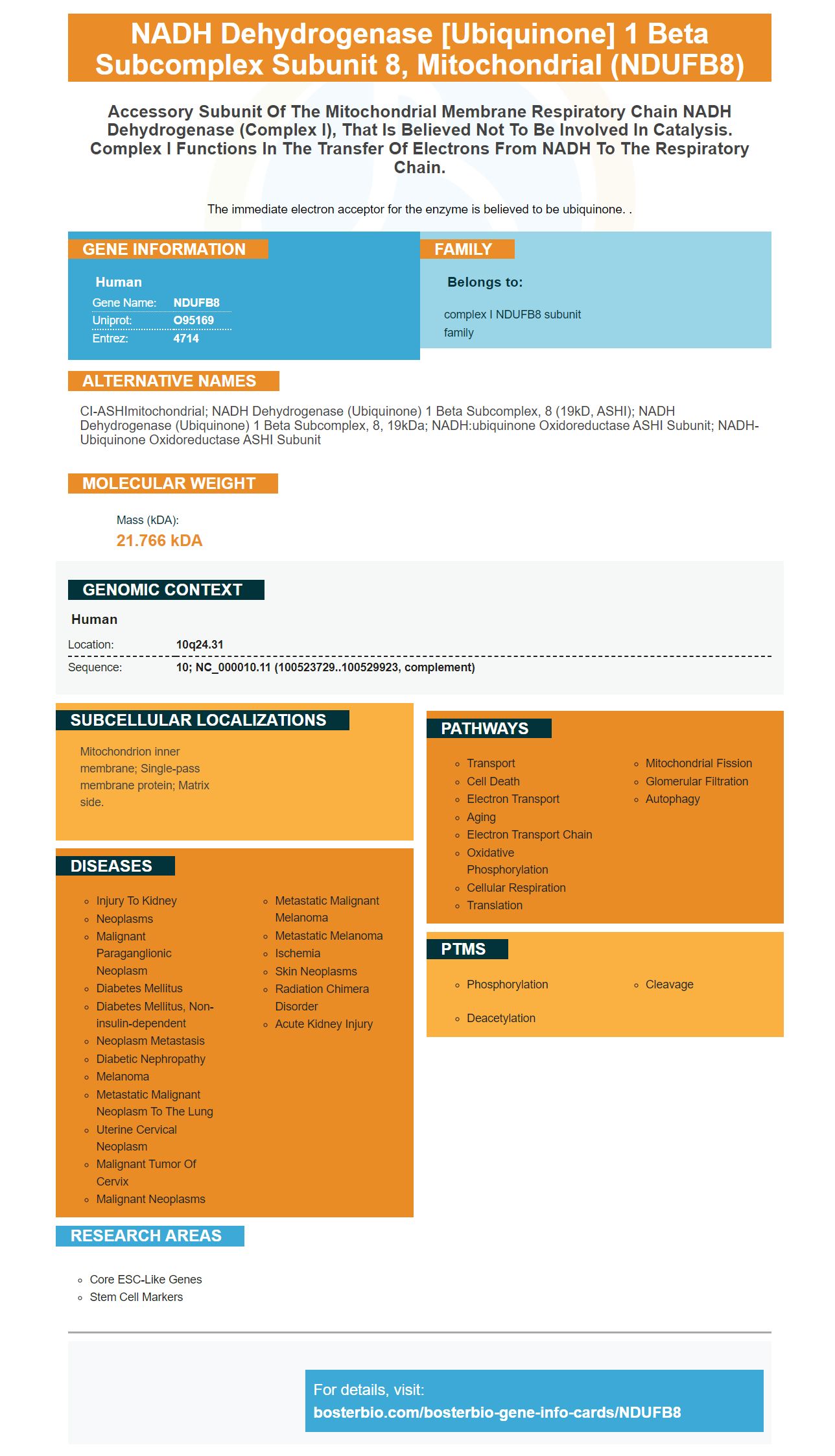

Facts about NADH dehydrogenase [ubiquinone] 1 beta subcomplex subunit 8, mitochondrial.

The immediate electron acceptor for the enzyme is believed to be ubiquinone. .
| Human | |
|---|---|
| Gene Name: | NDUFB8 |
| Uniprot: | O95169 |
| Entrez: | 4714 |

| Belongs to: |
|---|
| complex I NDUFB8 subunit family |

CI-ASHImitochondrial; NADH dehydrogenase (ubiquinone) 1 beta subcomplex, 8 (19kD, ASHI); NADH dehydrogenase (ubiquinone) 1 beta subcomplex, 8, 19kDa; NADH:ubiquinone oxidoreductase ASHI subunit; NADH-ubiquinone oxidoreductase ASHI subunit
Mass (kDA):
21.766 kDA

| Human | |
|---|---|
| Location: | 10q24.31 |
| Sequence: | 10; NC_000010.11 (100523729..100529923, complement) |
Mitochondrion inner membrane; Single-pass membrane protein; Matrix side.


You've probably heard of NDUFB8 Marker if biology is your major. But you may not know who it is. Below are some details about Boster Bio's history and applications. This information should help to decide if this marker is right. If not, you can continue reading to learn about the inventor.
The history of NDUFB8 markers is varied. It has been used in studies on cells taken from Parkinson's patients and healthy cells. Researchers discovered that cells with elevated NDUFB8 levels had higher lewy bodies than healthy cells in one study. However, a subsequent study revealed that the marker is highly expressed in cells suffering from a rare genetic disorder called aifm1 deficiency.
We have identified the primary antibody and secondary antibodies for NDUFB8 markers using an optimised sequencing. TOMM20, which is a well-established marker to measure mitochondrial mass, was used for probes of the NDUFB8 markers. Pan-cytokeratin antibody and DAPI were used to normalize immunofluorescence signals. Despite using lower concentrations for other markers, the optimal fluorescence intensity was maintained.
The MTCO1 - NDUFB8 staining sequence was placed prior to NDUFB8. This reduces the possibility of reducing antigen. The a-TOMM20 antibody was placed at position 2 between the MTCO1 and NDUFB8 staining cycles, providing additional heat-mediated denaturation step. The NDUFB8/HRP compound was then visualized using an Axioskop2 epifluorescence microscope fitted with a 40x objective. Images were captured using a DAPI or FITC filter.
Relative expression of NDUFB8 was compared between porin and a-synuclein in cells from patients with LBD. Researchers discovered that NDUFB8 was significantly greater than porin in cells bearing Lewy-body cells than neurons containing A-synuclein. Fig. 3. The NDUFB8 relative a porin analysis was also correlated with the presence of LBD patients having respiratory chain deficiencies.
Single cells contain many mitochondria. Tumour-rich tissue that has OXPHOS impaired will have higher levels mitochondrial biogenesis. Thus, the OD-NDUFB8 and MTCO1 marker can be normalised to mitochondrial mass markers OD-TOMM-20 to overcome bias due to differences in mitochondrial mass. The single-cell optical density data can then be normalized to the mitochondrial marker OD-TOMM-20 as well as the corresponding control data.
PMID: 9878551 by Loeffen J.L.C.M., et al. cDNA of eight nuclear encoded subunits of NADH:ubiquinone oxidoreductase: human complex I cDNA characterization completed.
PMID: 10570959 by Emahazion T., et al. Identification of 167 polymorphisms in 88 genes from candidate neurodegeneration pathways.


