This website uses cookies to ensure you get the best experience on our website.
- Table of Contents
1 Citations 1 Q&As
8 Citations
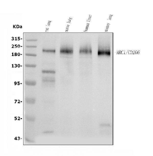
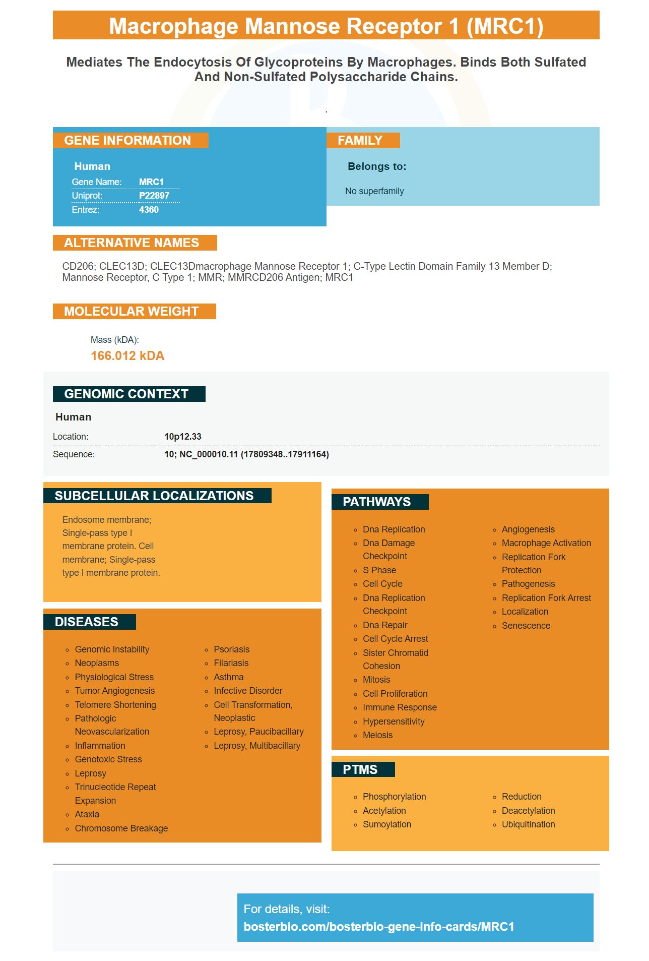
Facts about Macrophage mannose receptor 1.
.
Human | |
---|---|
Gene Name: | MRC1 |
Uniprot: | P22897 |
Entrez: | 4360 |
Belongs to: |
---|
No superfamily |
CD206; CLEC13D; CLEC13Dmacrophage mannose receptor 1; C-type lectin domain family 13 member D; mannose receptor, C type 1; MMR; MMRCD206 antigen; MRC1
Mass (kDA):
166.012 kDA
Human | |
---|---|
Location: | 10p12.33 |
Sequence: | 10; NC_000010.11 (17809348..17911164) |
Endosome membrane; Single-pass type I membrane protein. Cell membrane; Single-pass type I membrane protein.
This article will provide you with information about the company's history and the best uses for its MRC1 marker. You will also learn about Steven Boster, the creator of the marker, and the advantages of using it in your research. Also, you will gain an overview of the MRC1 monoclonal antibody. This antibody can be used to test samples of special species or to test applications. Additionally, you will learn a little bit about the MRC1 monoclonal antibodies.
Many companies offer anti MRC1 antibodies. These antibodies target the mannose receptor C type protein (also called CD206, CLEC13D or NIMA Kinase 2, akin to NIMA) and could have orthologs in yeasts monkeys, canines, and monkeys. These antibodies detect MRC1 protein in cells. They could also detect other proteins like CD163, which may be expressed on different cells.
Steven Boster, a smart businessman, earned a nickname in the lab due to his ability to develop hundreds of primary antibodies. After several years, he grew his company to become the largest catalog antibody company in China. Steven Boster also created PicoKine(tm) a proprietary ELISA platform in the late 1990s. This patent-pending technology allows for high sensitivity ELISA kits which are now available to researchers and biotech companies all over the world.
Many tissues and organs have high levels of the MRC1 gene. It is also found overexpressed on monocytes and dendritic cells, and is thought to play a role in adaptive and innate immune. The expression of MRC1 is evident by immunohistology on tissue macrophages as well as dendritic cell tissues. The antibody prevents the binding of glycoproteins and MR to the antibody, thereby labeling the cells.
This monoclonal antibody targets C-type family of lectins, 13 members D (CLEC13DL). It recognizes the mannose receptor. This type of receptor is expressed weakly at the cell surface, however the antibody may enhance staining after membrane permeation. The anti-mouse rat CD206 antibody clone MR5D3 was demonstrated to be effective in Western Blot and other immunohistochemical tests.
The mouse monoclonal antibody to recombinant human MRC1 is raised. This protein is located on the surface of many mammals. This monoclonal antibody can be diminished to a concentration which is suitable for your research. When making a monoclonal antibodies, ensure it is diluted to the working dilution of 0.02 percent sodium azide. The antibody is safe to use when it is diluted to 100 ug/ml.
The MRC1 monoclonal antibody targets macrophages M1 which are known to trigger and sustain inflammation throughout the body. The use of these antibodies can dramatically improve health by reducing activity of these immune cells. In actual fact, it is the most effective antigen against M1 macrophages. These cells are found in the bloodstream and play a crucial role in maintaining inflammation within the body.
PMID: 2373685 by Taylor M.E., et al. Primary structure of the mannose receptor contains multiple motifs resembling carbohydrate-recognition domains.
PMID: 2258707 by Ezekowitz R.A., et al. Molecular characterization of the human macrophage mannose receptor: demonstration of multiple carbohydrate recognition-like domains and phagocytosis of yeasts in Cos-1 cells.
*More publications can be found for each product on its corresponding product page