This website uses cookies to ensure you get the best experience on our website.
- Table of Contents
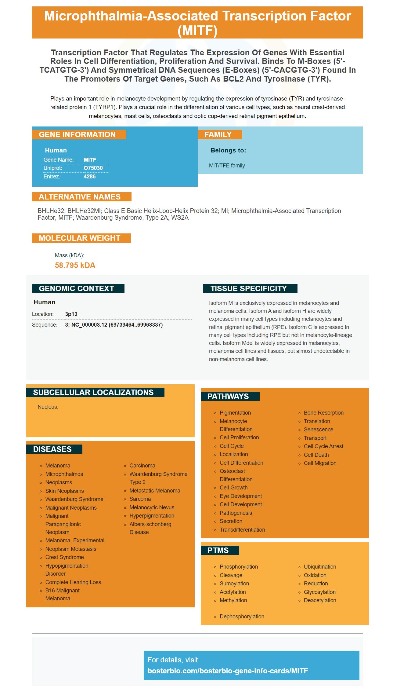
Facts about Microphthalmia-associated transcription factor.
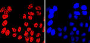
Plays an important role in melanocyte development by regulating the expression of tyrosinase (TYR) and tyrosinase-related protein 1 (TYRP1). Plays a crucial role in the differentiation of various cell types, such as neural crest-derived melanocytes, mast cells, osteoclasts and optic cup-derived retinal pigment epithelium.
Human | |
---|---|
Gene Name: | MITF |
Uniprot: | O75030 |
Entrez: | 4286 |
Belongs to: |
---|
MiT/TFE family |
bHLHe32; bHLHe32MI; Class E basic helix-loop-helix protein 32; MI; microphthalmia-associated transcription factor; MITF; Waardenburg syndrome, type 2A; WS2A
Mass (kDA):
58.795 kDA
Human | |
---|---|
Location: | 3p13 |
Sequence: | 3; NC_000003.12 (69739464..69968337) |
Isoform M is exclusively expressed in melanocytes and melanoma cells. Isoform A and isoform H are widely expressed in many cell types including melanocytes and retinal pigment epithelium (RPE). Isoform C is expressed in many cell types including RPE but not in melanocyte-lineage cells. Isoform Mdel is widely expressed in melanocytes, melanoma cell lines and tissues, but almost undetectable in non-melanoma cell lines.
Nucleus.
This article will discuss Boster Bio's Anti-Microphthalmia Transcription Factor Monoclonal Antibody (MITF) and its application to flow cytometry. We'll also discuss ISH Primers for lncRNAs GLIS2-AS1 & PV1.
The Boster Bio Anti-Microphthalmylia Transcription Factor MonocloNeo antibody binds to the symmetric DNA sequences of target genes to regulate gene expression. It plays a crucial role in the differentiation and maintenance of different cell types. It has been validated using multiple methods. This monoclonal antibody was designed to target mutated MITF.
This antibody recognizes MITF-antigens from rat and mouse. It is approved for use in Western Blot as well as flow cytometry. It has been tested in Flow Cytometry and ELISA. It is highly recommended to detect mtf related proteins.
The antibody recognizes both short and long forms. The melanocytes express the short form in two bands at 52kDa. The long form is found in osteoclasts. This antibody is sensitive to both types mi proteins and is very specific.
The results of the current trial with the Boster Bio Anti-Microphthalmic Antibody have been welcomed by the immunology community. This antibody is derived using a cloned lymphocyte from patients with metastatic melanomas. This research is a significant step forward in treating melanoma. It has also helped us understand how the disease develops.
Researchers from Japan have shown that the treatment of a melanoma involving the OX40 gene is effective against a wide range of diseases. In this study, the antibody reduced the number of tumor-causing T cells by increasing the number of regulatory CD4+ T cells in the patient. Boster Bio Anti–Microphthalmia Transcript Factor Monoclonal Antibody also reacted to tumor-lysed dendritic cell-pulsed dendritic cell samples in a similar study.
Mitf is key to regulating TRP-1 expression. It binds directly to the "Mbox" region of the promoter. This binding can trigger TRP-1 transcription. Mice with a mitf gene knockout exhibit 13 different pigment-related defects, including eye and coat color defects. Mitf is also involved during early T-cell development. Early T-cell development begins in the bone, where hemitopoietic precursor cells differentiate along various progenitor lines.
Autophagy is a highly conserved and self-degrading process that plays a critical role in cellular survival. Autophagy is dependent on tumor growth for many reasons, including chemoresistance or PDA development. PVT1 also plays an important role in PDA development. ULK1, an amino acid- and energy-regulated molecule, can trigger autophagy.
Primer sequences for lncRNA GLIS2 and lncRNA PVT1 were determined by PCR. To amplify GLIS2-AS1 or PVT1, total DNA was extracted from Jurkat cells using a PARIS Kit by Life Technologies. GAPDH, U6 and PVT1 were used as markers to standardize the expression of b-actin. DCt was used to analyze the amplification products.
ISH primers for lncRN PVT1 and GLIS2-AS1 were also developed to detect lncRNA expression within cells. These RNAs are commonly expressed in tumor cells. PVT1 interacts with several proteins, including c-Myc and the proliferation-associated nucleolar protein NOP2. Additionally, HULC regulates PRKACB expression in liver cancer cells.
The differential expression of lncRNAs PVT1 or GLIS2-AS1 in human parathyroid tumors is seen. These RNAs are potential markers in PCa or PAd, according to ISH. These data suggest that these two genes may be associated to multiple cancer features. Next is to test for cancers using lncRNAs and to evaluate the impact of circulating levels on various organs.
These lncRNA markers can be used to distinguish between cancers, as demonstrated by ISH-PCR tests. This approach can distinguish between tumors without and with the CDC73 mutation. PVT1 was also shown to be more sensitive than GLIS2-AS1 for detecting prostate carcinoma in prostate tumors.
To perform ISH a sense and antisense plasmid was transfected into human NPC cell lines. We used the BLOCK iT RNAi designer (THR) as well as Pierce(tm), RNA 3' End Desthiobiotinylation kit from Thermo Fisher Scientific for the ISH. These cells were then transfected with shFOXD3AS1 and scramble controls plasmids.
LncRNA GLIS2-AS1 (FOXD3-AS1) and FOXD3 -AS1 are other lncRNAs that have cancer-related roles. FOXD3-AS1 is overexpressed in NPC, despite having no protein-coding function. Based on GSE Database data and TCGA samples of Head & neck squamous Cell carcinoma, the relative expressions of these two lncRNAs can be determined.
SPSS 17.0 software (SPSS, Chicago) was used for statistical analysis. In cases with multiple samples, all numerical data were presented as means + standard deviation (M–SD). Non-parametric variables can be analyzed with Student's t and chisquare tests. Parametric variables were analysed using one-way analysis. To determine correlation, Pearson rank correlations were used. We also used the Kaplan-Meier method to determine survival.
There are many uses for flow cytometry. It has many benefits that range from studying the immune system to treating cancer and other infectious diseases. This powerful tool can analyze mixed populations of cells and allow for quick, quantitative analysis. The ability to sort live cells also allows researchers to monitor the immune response to a variety of illnesses. The results are sorted by size and antigenic marker to allow you to see them. The flow cytometry results are then used in research methods.
Flow cytometry is used to count and analyze cells that have a physical feature or marker. Electrostatic cell sorting is one of the most important applications of flowcytometry. This method separates cells according their subtype or expression level of a specific epitope. FACSTM analysis is this process. The data collected by flow cytometry can be analyzed to determine if a cell is cancerous or not.
The most common application of flow cytometry is immunophenotyping. Immunophenotyping involves staining cells by antibodies that are specific against antigens on cell surfaces. Monoclonal antibody are used to target these antigens. The Human Leukocyte Differentiation Workshops gives "cluster of differentiation" numbers. For instance, antibody CD3 - a coreceptor to T cells - is designated as cluster number 3
Fluorescent dyes are also used. Fluorescent dyes are molecules that absorb light energy at one wavelength and emit it at another, usually in nanoseconds. These properties make flow cytometry possible. There are many options for fluorescent dyes, making it easy to find the right reagent. They are applicable to many fields of research. Flowcytometry can be used to analyze samples.
In modern flow cytometry, fluorescently conjugated antibodies are used to label specific structures on cells. A laser beam interacts to fluorescing cells and creates a pulse of photon emissions over a time period. The PMT detects this pulse, and the voltage that is produced directly correlates with fluorescence intensities. The instrument can detect many channels if fluorescence intensity is high.
Applications of flow cytometry include the study of single particles and cells. This method analyzes single particle physical properties to measure their properties. The fluidics method is vital for studying single cells. It prevents the sample fluid from smearing in three dimensional space and prevents it mixing with the sheath fluid. The fluidics system also prevents the nozzle from blocking or mixing with the central chamber fluid.
Extensive research has been done on nanoparticle based applications. This technique is only possible if we have a clear understanding of nanoparticles' fate in cellular environments. It has been difficult to find analytical methods that can detect and quantify nanoparticles without fluorescent properties, or those that contain special elements. A recent study used flow cytometry to determine in situ NPs from mammalian cells. QPM is uncompatible with high-throughput flucytometry due its low camera frame rates.
PMID: 9647758 by Amae S., et al. Identification of a novel isoform of microphthalmia-associated transcription factor that is enriched in retinal pigment epithelium.
PMID: 8069297 by Tachibana M., et al. Cloning of MITF, the human homolog of the mouse microphthalmia gene and assignment to chromosome 3p14.1-p12.3.