This website uses cookies to ensure you get the best experience on our website.
- Table of Contents

10 Q&As

1 Citations 16 Q&As
1 Citations 4 Q&As


Facts about Tyrosine-protein kinase Mer.

Following activation by ligand, interacts with GRB2 or PLCG2 and causes phosphorylation of MAPK1, MAPK2, FAK/PTK2 or RAC1. MERTK signaling plays a role in various processes such as macrophage clearance of apoptotic cells, platelet aggregation, cytoskeleton reorganization and engulfment.
| Human | |
|---|---|
| Gene Name: | MERTK |
| Uniprot: | Q12866 |
| Entrez: | 10461 |

| Belongs to: |
|---|
| protein kinase superfamily |

c-Eyk; c-mer proto-oncogene tyrosine kinase; C-mer; EC 2.7.10; EC 2.7.10.1; MER receptor tyrosine kinase; Mer; MerTK; MGC133349; Receptor tyrosine kinase MerTK; RP38Proto-oncogene c-Mer; STK kinase; tyrosine-protein kinase Mer
Mass (kDA):
110.249 kDA

| Human | |
|---|---|
| Location: | 2q13 |
| Sequence: | 2; NC_000002.12 (111898607..112029561) |
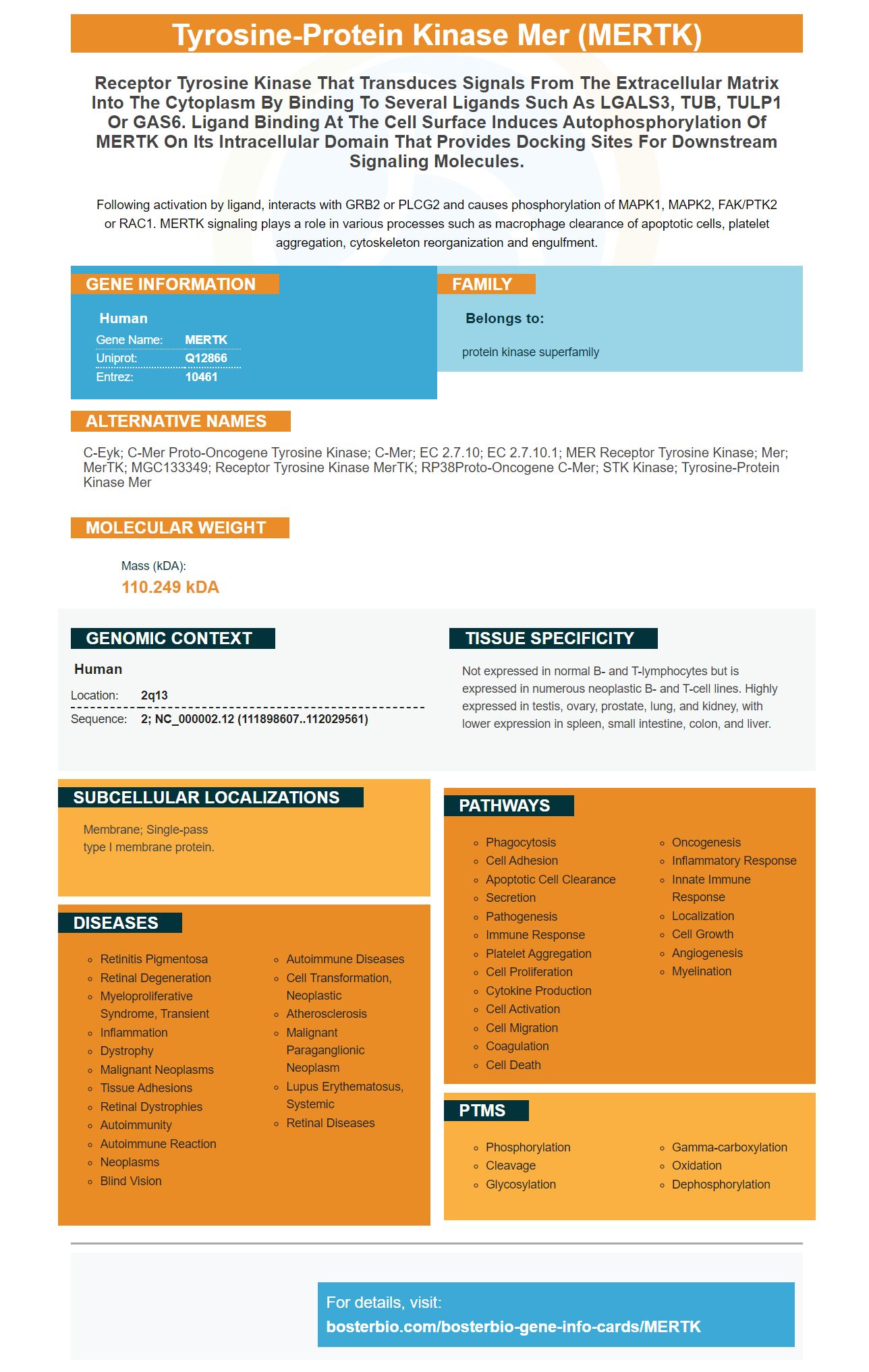
Not expressed in normal B- and T-lymphocytes but is expressed in numerous neoplastic B- and T-cell lines. Highly expressed in testis, ovary, prostate, lung, and kidney, with lower expression in spleen, small intestine, colon, and liver.
Membrane; Single-pass type I membrane protein.


MERTK is a transmembrane proteins that is involved in the process of efferocytosis. It may rely on the RAF pathway to allow it to metastatically expand. It is also a crucial marker for immune cells in mice and humans. In this article, we'll examine the most effective uses of the MERTK marker. Read on to learn more!
The CD14thCD16th monocyte population had the highest levels of MERTK expression in the study described. This suggests that therapies aimed at enhancing innate immune responses could prevent secondary infections. Strategies to boost sMER activity may be a novel immunotherapeutic approach. MERTK could be used to bring back the innate reaction to a microbial threat and could be used as an immunotherapy option for ACLF patients.
The LINE-1 element has been added into the MERTK gene. The element is located in the same way as MERTK gene. The expression of the corresponding mRNA can be increased by inserting LINE-1 in the MERTK genes similarly to MERTK. Further studies are needed to determine the degree of expression for MERTK in affected Swedish Vallhunds.
The expression of MERTK in monocytes from ACLF patients is increasing. MERTKth monocytes preferentially migrate across endothelia , and eventually into the liver. UNC569 treatment decreased inflammatory monoocytes expressing cytokine expression. Patients with ACLF have seen their survival rates and incidences of ACLF decreased by monocytes expressing MERTKth.
PRA is a broad term that refers to progressive retinal diseases in dogs. The Swedish Vallhund breed recently found a new type of disease. The gene MERTK was discovered on the canine chromosome 17. The analysis of expression of the affected retinas of Vallhunds revealed that the MERTK gene expression was six times greater than the expression of healthy controls. The association between MERTK and PRA in the affected Vallhunds was confirmed in UK Swedish Vallhunds. Using whole genome sequencing, researchers sought to identify the causal variant underlying the relationship.
Researchers were able to detect macrophages that express MERTK using confocal microscopes. In this study, liver tissue from ACLF and orthotopic patients undergoing liver transplantation was collected. For normal control tissue, liver margins from resections were used as controls. The immunohistochemistry method was also used to identify MERTKthCD68th MERTKthCD163th expressions in ACLF patients.
In cancer, efferocytosis is an important process that removes cells that are apoptotic from the body. Efferocytosis disturbances can result in cytosolic leakage and tissue exposure to toxic substances. Efferocytosis dysfunction can also cause the induction or maintaining of canonical autophagy within cells adjacent to each other.
Recent research suggests that there is a link between efferocytoses and cancer progression. This process gets more intense in the postpartum time frame when it allows for the inflow of healing microphages. This is a sign of metastasis, cancer progression and can be attributed to efferocytosis. It suggests that anti-tumor treatments could be more efficient when they target efferocytosis.
As cancer therapy targets apoptotic cell to eliminate them, the removal of Apoptotic cells requires coordination between efferocytosis and LC3-associated phagocytos. In addition, tumor cells utilize anti-inflammatory and immunosuppressive signals created by LC3-associated phagocytosis. This is a major factor in the advancement of the disease. However, mice lacking LAP demonstrated more rapid growth of tumors, which point to the significance of regulators of LAP in controlling the phagocytosis process.
Utilizing a GEO database, researchers looked at the RNA-seq information in a mouse osteosarcoma tumor cell line and normal tissue to determine whether a tumor is expressing MerTK. The expression of MerTK was significantly higher in osteosarcoma tissues than normal tissues. In osteosarcoma, tumor cell, macrophages and fibroblasts are the most abundant cells. Researchers compared the levels of merTK mRNA expression in the K7M2 cancer cell line, RAW264.7 macrophage cell line and NIH-3T3 cell fibroblasts.
The activation of Mertk activates the MEK ERK-Tpl2-NFkB complex, which blocks conventional NFkB signaling. These changes lead to alteration of macrophage response without Polarization. Similar to the previous increase in the expression of NFkB and p50 have been found to alter macrophages that respond LPS. Mertk is essential for the control of immunity.
A low response to radiation therapy has been associated with a lack of Mertk in the mouse model for cancer. However, in mice with Mertk tumor growth is decreased. Wild-type mice exhibit a transient level of control followed by outgrowth. Mertk-deficient mice, on contrary, can remain alive after complete removal of tumors. This is crucial in the treatment of cancer.
Although it isn't clear what mechanisms members of the RAF family drive metastasis, it is believed that CRAF stimulates autocrine TF-b release as well as activation of ERK/MAPK pathways. ARAF is also involved in the process of tumor formation. The TGF-b signaling pathway is thought to be a major cause of prostate cancer bone metastasis.
Mek1/2 and Rac are protein kinases. They MEKs activate Raf by phosphorylation and binding of residues at the activation segment of the kinase domain. MEK activation is also controlled by RAF kinases. Mutated MEK reduces cytokine-dependent haematopoietic cell development and triggers modifications in the morphology of NIH-3T3 cell.
These results suggest that RAF family members have the capacity to enhance metastatic capability in different types of tumors. We now know that RAF members drive bone metastasis, and NTRK2 and MERTK can promote metastasis in the lungs and viscera. Therapeutic interventions that target the RAF family are an essential component of cancer research. Our findings suggest that MERTK could be a great candidate for cancer drugs that block the RAF pathway.
We also know that activating Raf can cause resistance to drugs. This resistance is connected to the inhibition of the drug-pump Mdr-1 and Bcl-2. We can reduce resistance to drugs used in metastatic therapy by blocking these pathways. The RAF pathway is also involved in other functions in the field of cancer biology. Raf is activated by kinases which cause resistance to chemotherapy. Metastasis may also be prevented by blocking RAF.
MERTK is a gene found on the surface of numerous immune cells of mice and humans. It is thought to be involved in the regulation of the T cell response as well as the innate effector function. When activated in the immune system, MerTK increases the number of T cells, including DCs and macrophages. This effect is not completely understood. A molecule called PROS1 regulates MERTK.
In the present study, MERTK expression was examined in purified CD4+ T cells that were cocultured with mouse DCs in the presence of CD28 beads that a-CD3 were present. T cell growth was stimulated by DCs that were with naive CD4+ T cells and a-MERTKmAb. Additionally, DC maturation status did not affect the results.
Three different panels of markers were utilized to test the expression of MERTK. In the first panel, gene/protein combinations were evaluated on human T-cell markers Cd5 (CD5), Cd4a (CD8), and Il2ra (CD25) In the second panel, markers for mouse myeloid cells comprise Ly6g (Ly-6G) and Fcer1g CD64/F4/80.
While the expression of MERTK is upregulated in DCs in vitro, it is not clear if the protein is enhanced in the presence dex. Studies have revealed that DCs treated (MDA-IVD) exhibit high levels of. Furthermore, MERTK was upregulated in cells that are not yet naive, suggesting that dex treatment can boost DC growth.
Expression of MERTK can be observed in the immune infiltrates of breast cancer tumors. It is commonly found in the presence of mononuclear cells. However, MerTK expression was also observed in small round mononuclear cell types in the tumors as well as healthy breast tissue that is adjacent to them. MERTK can also be detected in healthy breast tissue but this is rare. In addition to the melanoma, MerTK expression is also found in breast cancer.
PMID: 8086340 by Graham D.K., et al. Cloning and mRNA expression analysis of a novel human protooncogene, c-mer.
PMID: 11062461 by Gal A., et al. Mutations in MERTK, the human orthologue of the RCS rat retinal dystrophy gene, cause retinitis pigmentosa.
*More publications can be found for each product on its corresponding product page


