This website uses cookies to ensure you get the best experience on our website.
- Table of Contents
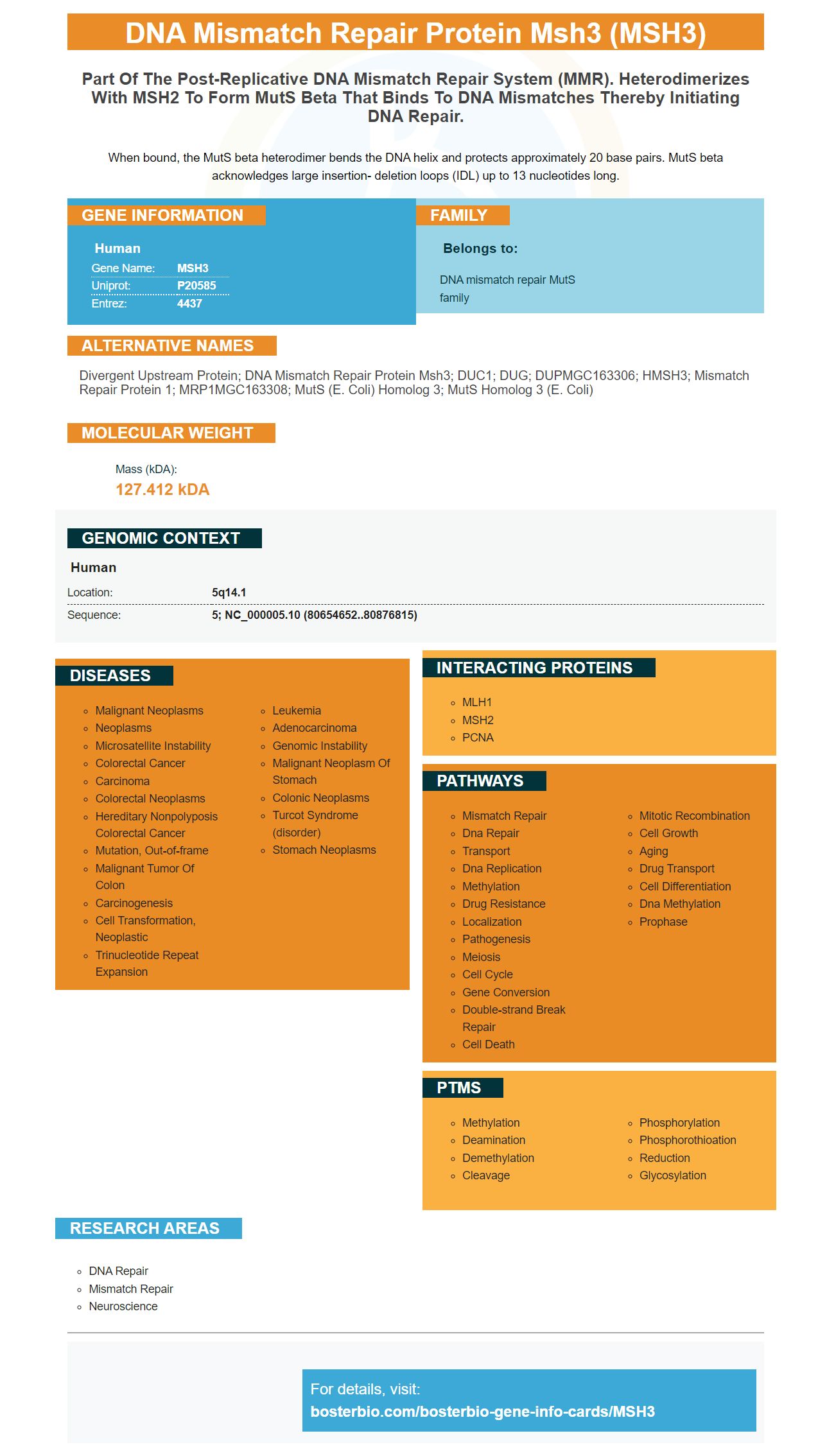
Facts about DNA mismatch repair protein Msh3.
When bound, the MutS beta heterodimer bends the DNA helix and protects approximately 20 base pairs. MutS beta acknowledges large insertion- deletion loops (IDL) up to 13 nucleotides long.
Human | |
---|---|
Gene Name: | MSH3 |
Uniprot: | P20585 |
Entrez: | 4437 |
Belongs to: |
---|
DNA mismatch repair MutS family |
Divergent upstream protein; DNA mismatch repair protein Msh3; DUC1; DUG; DUPMGC163306; hMSH3; Mismatch repair protein 1; MRP1MGC163308; mutS (E. coli) homolog 3; mutS homolog 3 (E. coli)
Mass (kDA):
127.412 kDA
Human | |
---|---|
Location: | 5q14.1 |
Sequence: | 5; NC_000005.10 (80654652..80876815) |
In this article, we'll review how the MSH3 Marker is used in a variety of biological assays, including Western Blot, ELISA, and Flow Cytometry. To help you decide if this is the right biological marker for your experiments, we've outlined the most common uses of the MSH3 Marker. This article will also discuss the advantages and disadvantages of these various assays.
MSH3 is a highly conserved sequence found on the genome. It is an essential component of the MMR pathway, a DNA repair system that corrects replication errors and deletes or insertions in the daughter strand of DNA. It is crucial to maintain genomic stability, but defects in this pathway can lead to severe disease, including Lynch syndrome. To learn more about the role of MSH3, read on!
MutS and MutL homologs are proteins that form heterodimeric complexes. The MSH2--MSH3 complex is partially redundant and functions in base-base mispair repair, small insertion/deletion mispair repair, and recombination. It also plays a limited role in larger insertion/deletion mispair repair.
Mutations in mlh3 result in a defect in the repair of insertion/deletion mispairs, mononucleotide-run replication errors, and mismatch repair. Mutations in mlh3 cause an increase in frameshift mutations in both mutants and plasmid strains lacking MSH6. The double mutant, mlh3 pms1, had a higher mutation rate than the single mutant. The mutations in mlh3 pms1 caused defects in MSH3-dependent repair.
MSH3 is used in various cancer diagnostics. MSI phenotype is characterized by loss of MLh2 protein expression and hypermethylation of MLh2 gene promoter. Patients with Lynch syndrome have increased risk of developing colorectal, biliary tract, and brain tumors. However, there are other tumors that can occur despite MSI.
The MSH3 ELISA kit from Boster Bio is an ideal choice for researchers looking to quantify MSH3 levels in human tissues and sera. The company manufactures polyclonal antibodies that are optimized for IHC, WB, and Flow applications. Their antibodies have undergone rigorous validation against a panel of 250 tissue samples. Additionally, they have been tested against untransfected cell lines and other recombinant proteins.
The MSH3 ELISA kit from Boster Bio uses a rabbit anti-MSH2 antigen as the primary antibody and a goat anti-rabbit IgG-HRP as the secondary antibody. Samples were loaded into the sample wells under reducing conditions. Proteins were transferred to the nitrocellulosmutembrane at 150 mA for 50-90 minutes. Finally, samples were blocked with 5% non-fat milk/TBS for 1.5 hours.
In this study, we characterized the effect of MSH3 gene silencing on the metabolic processes of cells. This marker represses several proteins involved in cell metabolism, including asparagine synthase, biliverdin reductase A, and NADH dehydrogenase, an important electron transport chain enzyme. We also examined the MSH3 protein in the plasma to study the effects of MSH3 on cell cycle progression.
The MSH3 gene is essential for cancer development, as its absence is linked to a defect in the immune response in MSI-H CRCs. It also participates in homologous recombination and the repair of DSBs. When this gene is missing in cancer cells, DSBs accumulate in the cell's genome, resulting in aneuploidy and poor prognosis.
In human cancers, MSH3-deficiency results in DSBs and EMAST, a mutator phenotype that promotes DNA-damage responses. The MSH3 marker was first discovered in colon cancer and subsequently found in non-small cell lung cancer. In mice, however, MSH3 silencing is not sufficient for EMAST to occur, despite its prognostic value.
In addition to the MSH3 gene, the MSH6 gene is also important. MSH6 is a tember of the MutSa family, and has a role in regulating the stability of tetranucleotide repeats. Loss of MSH3 in cancer cells leads to lower levels of MSI in the cell. In addition to the MSH3 marker, other cellular markers such as EMAST or EMA have also been identified.
Other proposed markers for cancer cell differentiation are CDX2 and MSH3. These three markers are also being investigated. Several more markers are being developed for future research. These include the MSH3 marker, EMAST, and the proposed markers for immune cell differentiation. It is important to note that the revised Bethesda panel includes three mononucleotide markers. Flow Cytometry with the MSH3 marker is an efficient way to determine cell viability in cancer patients.
The MSH3 gene is a unique MMR protein. It has been found that the repression of MSH3 results in significant changes in the expression of 202 proteins involved in fundamental cellular pathways. MSH3 deficiency is associated with an upregulation of the 26S proteasome regulatory subunit 7 (RPC) gene, which is involved in the ubiquitin-degradation process.
This study uses an antibody called MSH2 to detect MSH2 expression in the human body. The antibodies were purchased from Boehringer Mannheim and prewashed before use. The antibodies were used for immunoprecipitation and Western blot analysis. The antibody detected Histone H3 at a 17 kD molecular weight. The antibodies detected the protein only in the presence of ATP.
The immunohistochemistry analysis revealed a negative association between MSH2 and MLh2, but no evidence of causality. Both the MSH3 and MMR proteins play minor roles in human tumorigenesis. It is important to distinguish the differences between the two proteins so as to improve our ability to intervene in CAG/CTG expansion. However, further studies need to be conducted to determine the exact role of these proteins in tumorigenesis.
Antibodies for PCNA and MSH3 are monoclonal and polyclonal in nature. Both proteins react with PCNA and MSH3 in animal samples. Boster Bio uses rabbit and mouse as the models for its antibodies. The protein is an auxiliary protein of DNA polymerase delta that enhances the polymerase's processibility during elongation of the leading strand. PCNA is known to stimulate 3'-phosphodiesterase, but not AP endonuclease.
MSH2 and MSH3 are a heterodimer that recognizes mismatch repair. The hMutLa complex of MLh2, PMS2, and PCNA restores the function of mismatch repair in colon tumors. The PRCA gene, which encodes PCNA, has been identified as a participant in mismatch repair. In addition, p21 is a universal cyclin-dependent kinase inhibitor and inhibits strand-specific mismatch repair.
The 3.2 kb marker shows the original location of the strand-specific mismatch or strand break. It also shows the location of the oligonucleotide probe used in the experiment. 6.4 kb is mapped for ligated substrates and normal repair products. The 3.2 kb 5' of Bsp106 represents the heteroduplex strand-break and MSH3 marker in Boster bio.
PMID: 2722860 by Fujii H., et al. Isolation and characterization of cDNA clones derived from the divergently transcribed gene in the region upstream from the human dihydrofolate reductase gene.
PMID: 8942985 by Acharya S., et al. hMSH2 forms specific mispair-binding complexes with hMSH3 and hMSH6.