This website uses cookies to ensure you get the best experience on our website.
- Table of Contents
Facts about Major histocompatibility complex class I-related gene protein.
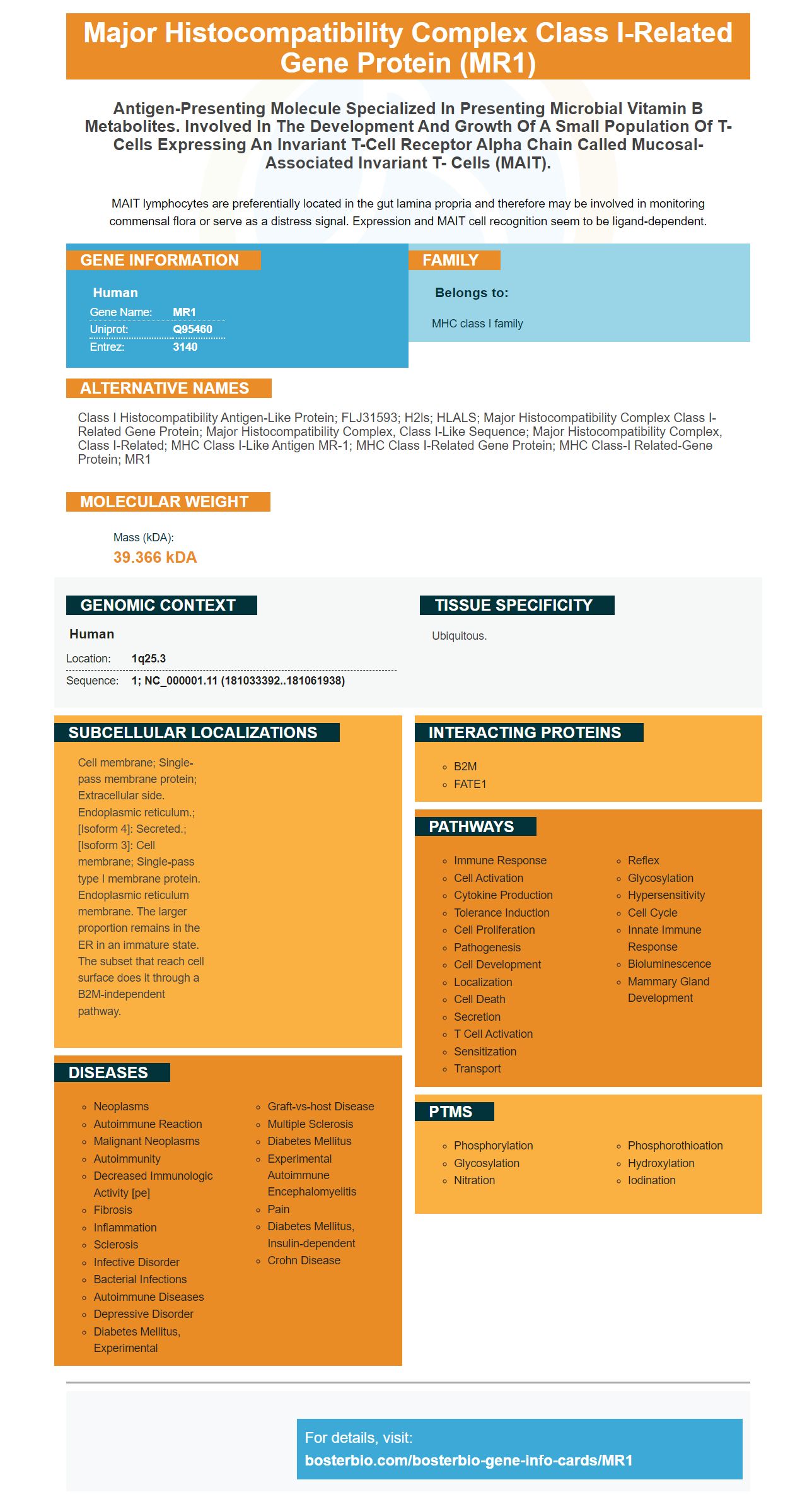
MAIT lymphocytes are preferentially located in the gut lamina propria and therefore may be involved in monitoring commensal flora or serve as a distress signal. Expression and MAIT cell recognition seem to be ligand-dependent.
Human | |
---|---|
Gene Name: | MR1 |
Uniprot: | Q95460 |
Entrez: | 3140 |
Belongs to: |
---|
MHC class I family |
Class I histocompatibility antigen-like protein; FLJ31593; H2ls; HLALS; major histocompatibility complex class I-related gene protein; major histocompatibility complex, class I-like sequence; major histocompatibility complex, class I-related; MHC class I-like antigen MR-1; MHC class I-related gene protein; MHC class-I related-gene protein; MR1
Mass (kDA):
39.366 kDA
Human | |
---|---|
Location: | 1q25.3 |
Sequence: | 1; NC_000001.11 (181033392..181061938) |
Ubiquitous.
Cell membrane; Single-pass membrane protein; Extracellular side. Endoplasmic reticulum.; [Isoform 4]: Secreted.; [Isoform 3]: Cell membrane; Single-pass type I membrane protein. Endoplasmic reticulum membrane. The larger proportion remains in the ER in an immature state. The subset that reach cell surface does it through a B2M-independent pathway.
If you're new to staining mRNA, you might be wondering what the best method is. Boster Bio offers two main systems: the primary and secondary ABC. Both systems validate antibodies against positive and negative samples for high affinity and specificity. Boster also rewards reviewers who are early in their reviews with product credits. The system is accessible to all scientists in the world. Below are some of the most popular uses of the MR1 marker.
The identification of proteins in biological samples is not always straightforward. In many cases, the use of a primary-secondary-ABC system allows researchers to easily identify proteins in their specimens. The system utilizes avidins that have been conjugated to signal molecules like Horseradish Peroxide (HRP). The detection system is extremely sensitive and specific and has exceptional low-background and high-specificity properties. Similar systems have been created using organic polymers and polysaccharides. Boster Bio's Super Vision Detection Kits are an outstanding example of this revolutionary approach.
Boster Bio's Super Vision detection kits are among the most recent developments in the field of protein detection. These kits employ avidins that are coupled to signal molecules like Horseradish Peroxide to identify and measure proteins. They have high specificity and low background. Boster's Super Vision Detection kits are a great example of innovative user-friendly detection systems.
MR1-restricted T cells recognize a specific cancer metabolite , which has distinct TCRs and , therefore, offer an untapped potential for pan-cancer therapeutic agents. In this article, we will look at the physiology and function of MR1-restricted cells and how they can be used in cancer immunotherapy. It's important to note that cytotoxic T cells that recognize a specific metabolite are referred to as MR1-restricted T cells.
The presence of a protein called CD1a in human MR1-restricted MR1 T cells is an indication that they are human. CD1b-specific T cells are more rare and can be easily identified by staining CD1b with CD1b Tetramers. The molecule binds with an antigen that has the same peptide sequence of CD1a.
Neonatal BCG vaccination has increased the number of CD4+CD26+CD161+T cells frequencies that expressed IFNg but they did not express the MAIT TCR Alpha gene segment. MAIT cells are traditionally classified as CD161+CD8+T cell, however, most studies were conducted on adults. The MR1-5 OPRU tetramer however, can detect CD4+CD161+ cell types, including those that express TRAV1-2.
Single-site mutants of the TCR are able to bind the lipids contained in CD1a. I72A, a single site mutant of the TCR all eleven lipids eluted by CD1a. This included DAG and triacylglycerol. This is the first time we have reported on a novel subset MR1-restricted MR1 T cells. This study offers insight into the TCR's response to a specific antigen.
It has also been established the phenotype for CD161++CD8a+ cells. They show high levels of CD161 and type-17 characteristics. These T cells are not only human, but there is increasing evidence that they are similar to CD161++CD8a+ CD8a+ TCRab+ cells and are, therefore, most likely to be deriving from them.
MR1-restricted cells are also identified in the Boster Bio database. Moreover they are implicated in a variety of diseases such as multiple sclerosis. But, despite the progress made, there are still many questions unanswered. Thus, identifying the precise role of these T cells in various diseases is essential to the development of vaccines.
MRI staining of MAIT cells and TRAV1-2 cells showed an overlap in their levels of IFNg. MR1-dependent and MR1-independent activation of MAIT cells is expected to be mutually beneficial. These cells also possess similar antimicrobial properties. However, the intracellular context is essential for the detection of MAIT cells. Therefore, MR1-5 tetramers could be a helpful tool for identifying MAIT cells.
MAIT cells have TCRs that are semi-invariant. TRAV1-2 and TRAJ33 are chains of TCRs that work together to facilitate the recognition of riboflavin derived Ags. MAIT cells also have TRAJ12, TRAJ20 and TRAJ33 in their repertoires. MR1 Tetramers were obtained from NIH tetramer facilities.
MR1-expressing T cells express the innate Va7.2+ T cells and show positive selection. The MAIT-a chain that is the canonical one was expressed by cells located on the gate's upper edge, whereas those at the lower edge showed different chains that were not MAIT-a. The MR1-positive cells can also serve as selector cells for the human MAIT cells of the thymus.
MR1 MAIT cells that are tetramer+ are largely CD8aa+ and DN. They lack CD8b however they do contain CD8aa homodimers. MR1 mait cells with tetramer exhibit the same phenotypes as conventional ab T cells. Therefore, intracellular transcription factor staining with the MR1 marker is helpful in identifying MAIT cells.
Utilizing this MR1-tetrameric protein as the activation marker, this technique was able to detect accurately activated MAIT cells. Mycobacterial studies showed that the CD69+CD26++ CD69-dependent population was always upregulated. Based on their MR1 dependence CD69+CD26++ may be used as an identifier for human MAIT cells.
Researchers use the MR1-tetramer to determine MAIT cells and to determine the mechanisms behind their activation. Conventional T cells react slowly when exposed to MHC-presented antibodies, whereas mucosal-associated invariant cells respond rapidly to non-peptidic substances. Utilizing the MR1 marker in MAIT cells provides the benefit of delving into the MAIT cell activation program and its effector responses. This technique is particularly useful in mycobacterial infections, where the MR1-mediated presentation of antibodies stimulates a variety of human CD8+ MAIT cell populations. Furthermore, MR1-mediated presentation of antigens to MAIT cells is thought to be a marker of combinatorial activation.
MAIT cells express tissue-homing regulators (Th27) and innate lymphoid marker markers, CD161 and CD218, which aid in identifying MAIT cell types. Similarly, MR1-expressing cell are identified by the expression of CD8a/B lectins. These cells also express markers related to the innate immune system, including MR1.
PMID: 7624800 by Hashimoto K., et al. A gene outside the human MHC related to classical HLA class I genes.
PMID: 11019920 by Parra-Cuadrado J.F., et al. A study on the polymorphism of human MHC class I-related MR1 gene and identification of an MR1-like pseudogene.