This website uses cookies to ensure you get the best experience on our website.
- Table of Contents
1 Citations 4 Q&As
Facts about Matrix metalloproteinase-24.
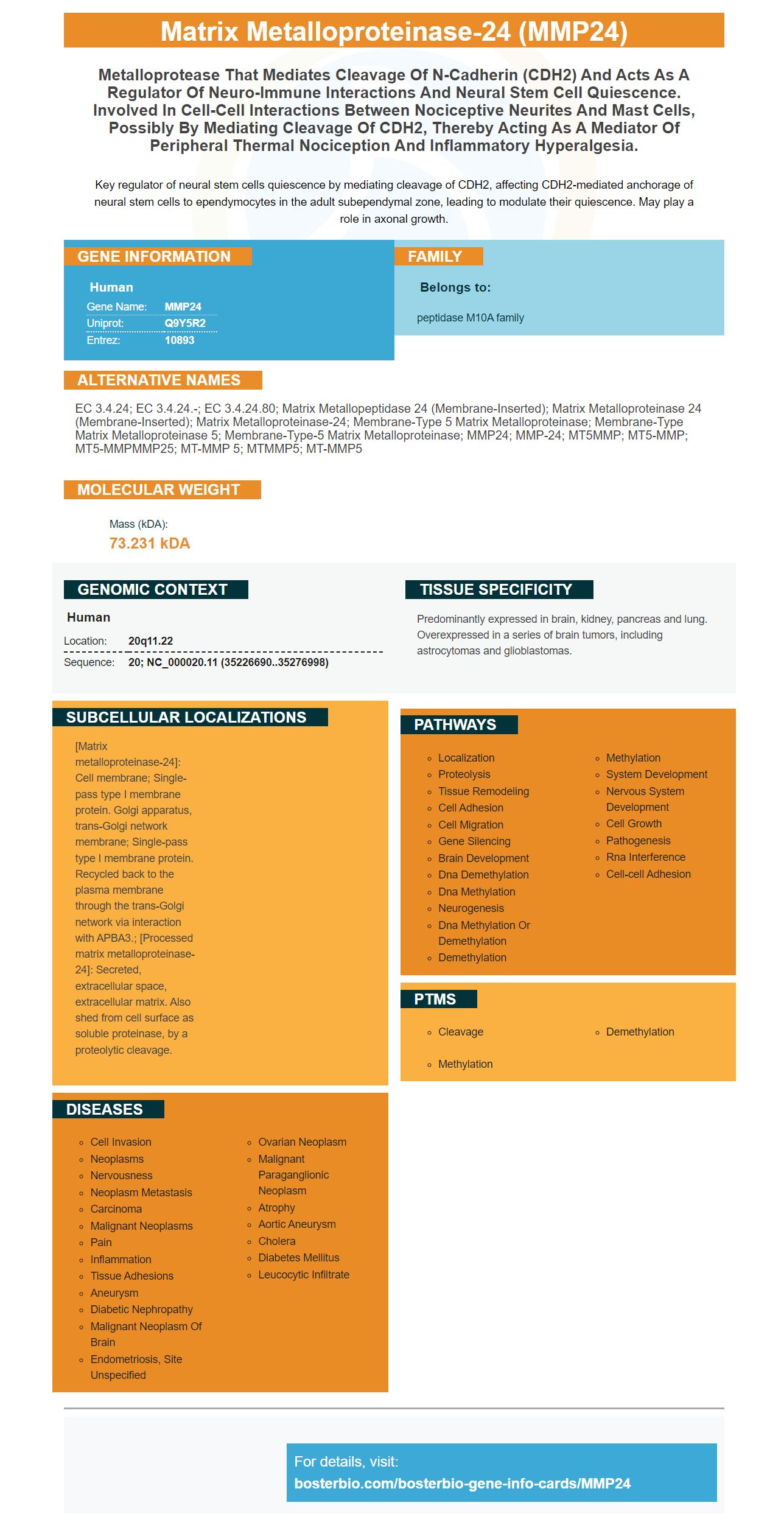
Key regulator of neural stem cells quiescence by mediating cleavage of CDH2, affecting CDH2-mediated anchorage of neural stem cells to ependymocytes in the adult subependymal zone, leading to modulate their quiescence. May play a role in axonal growth.
Human | |
---|---|
Gene Name: | MMP24 |
Uniprot: | Q9Y5R2 |
Entrez: | 10893 |
Belongs to: |
---|
peptidase M10A family |
EC 3.4.24; EC 3.4.24.-; EC 3.4.24.80; matrix metallopeptidase 24 (membrane-inserted); matrix metalloproteinase 24 (membrane-inserted); matrix metalloproteinase-24; membrane-type 5 matrix metalloproteinase; Membrane-type matrix metalloproteinase 5; Membrane-type-5 matrix metalloproteinase; MMP24; MMP-24; MT5MMP; MT5-MMP; MT5-MMPMMP25; MT-MMP 5; MTMMP5; MT-MMP5
Mass (kDA):
73.231 kDA
Human | |
---|---|
Location: | 20q11.22 |
Sequence: | 20; NC_000020.11 (35226690..35276998) |
Predominantly expressed in brain, kidney, pancreas and lung. Overexpressed in a series of brain tumors, including astrocytomas and glioblastomas.
[Matrix metalloproteinase-24]: Cell membrane; Single-pass type I membrane protein. Golgi apparatus, trans-Golgi network membrane; Single-pass type I membrane protein. Recycled back to the plasma membrane through the trans-Golgi network via interaction with APBA3.; [Processed matrix metalloproteinase-24]: Secreted, extracellular space, extracellular matrix. Also shed from cell surface as soluble proteinase, by a proteolytic cleavage.
You might be a beginner in the field. There are many factors that you need to take into consideration, including flow procedures and antibody titration. These are some tips and tricks that will help you optimize your experiments. Continue reading to learn even more. Here are some of our favorite uses for the MMP24 Marker. Find out why they're so useful.
The MMP24 gene encodes the protein that degrades extracellular matrix. It also breaks down other proteinsases. These proteins are involved in a range of normal physiological processes including tissue remodeling. Researchers can use the MMP24 mark to identify specific antibodies to an enzyme to determine if a particular protein is active. MMPs are part of the MMP family of enzymes, which is categorized as membrane-type.
Boster's MMP24 antibody recognizes it in multiple applications. It is a reagent used in WB applications. It reacts well with Rat, Mouse, Human. The antibody is stable at -20C for one year. Boster's MMP24 and primary antibodies are compatible. They are tested on a variety tissue types and species to ensure high specificity and affinity.
However, fluorescent IHC is used to detect multiple markers. Fluorescent mIHC is often used in conjunction with fluorescent dye-conjugated primary antibodies for extra amplification. Fluorescent mIHC can only be used with three markers because of the limitations of available filter sets. Additionally, it requires the use of primary antibodies raised against different species and isotypes. This method requires more than three markers to identify a target protein.
The secondary antibody binding substances are the basis of western blot antimatter detection methods. Boster Bio MMP24 Western blot antibodies depend on exposure to the secondary antibody, and ECL chemiluminescent blotting is the preferred method. This method can also serve as a tool in immunogenetics. It can detect proteins as low as 1ng within biological samples. This method is applicable to a wide variety of molecular biology applications, including immunogenetics.
Boster's MMP24 antibodies are tested on multiple platforms to verify their high specificity. Boster awards product credits to the first reviewer of the antibodies and rewards them with product credit. This program is open worldwide to scientists. All Boster antibodies have undergone rigorous validation for sensitivity and specificity. Boster also provides negative control for quality control to eliminate any nonspecific staining.
A weak western blot signal is an indicator of low or no protein. It can occur at any point in the process. It can also occur due to low levels of the antigen. It is important to use an antigen of another source in such cases to confirm the results. To break the quaternary structural, it is a good idea to heat the sample. Blurry bands can also occur from air bubbles within the transfer sandwich. To avoid this, make sure the transfer sandwich is properly prepared.
Western blots can be used to diagnose other than immunoblotting the protein of concern. A positive Western blot can confirm the diagnosis of Lyme disease or Alzheimer's disease. Moreover, it can also be used to detect proteins that are associated with a wide range of other diseases. It can also be used to confirm positive ELISA tests.
Chemiluminescent Western detect is a well-known method for research. It involves the use of an enzyme, known as enhanced chemiluminescence (ECL), to detect the presence of proteins. Luminol is a popular chemiluminescent substrate. Its half life is less than 1 minute. Enhanced chemiluminescence has a longer signal-to-noise ratio than Luminol.
The system includes a range of highly sensitive and robust reagents. ECL Prime is an example of this. Its stable signal emission allows for multiple exposures. This reagent is highly sensitive and has a very low background. Hybond ECL membranes offer excellent sensitivity with low background. Hybond ECL membranes are made of pure nitrocellulose and have excellent signal-to-noise ratios.
Researchers can detect protein presence using the same antibodies or different molecules using enhanced chemical luminescence. This method is the preferred choice of many experts because it has a high signal to noise ratio, high sensitivities, and a wide dynamic range. Consequently, it is ideal for researchers performing Western blot analysis. The enhanced chemiluminescent detection device has a wide range of uses.
The Enhanced Chemiluminescent substrate (ECL) is often used for Western blot applications. It reacts with HRP labeled antibodies to create luminescence. The amount of proteins present in the signal determines the intensity. This signal is used in indirect protein measurement. The intensity of the signal can be detected with a photographic film or imaging instrument. The ECL substrate agent is sensitive to long-term light. This should be avoided.
To prepare a gel to detect chemiluminescent light, wrap the blot membrane with clear plastic wrap or transparent plastic sheets protector. Place the membrane with the protein facing upwards. Next, expose the blot membrane to X-ray. The film should be exposed to the sun for one minute. The substrate should be exposed for about one minute. Light emission intensifies between five and thirty minutes after it is incubated. The resulting images are processed using a chemically luminescent detector and a photogrammetric camera.
The SITVue/DAB is an Intensification system that significantly enhances the chromogenic signals. It can be used in standard immunohistochemistry staining methods. Horseradish Peroxidase is used in this system to catalyze the enumeration of two separate Linkers. Then, the chromogenic signal is detected using a streptavidin-peroxidase conjugate and diaminobenzidine (DAB) substrate.
MMP24 is a protein which plays a key role in the degrading of collagen. In addition to its function in tissue repair, MMP24 also has numerous other uses. For example, in immunohistochemistry, MMP24 is used to identify fibroblasts and other inflammatory cells. It can also be used in cancer and infectious disease research. Boster Bio has high-affinity prima antibodies that are widely acknowledged by the research community. These antibodies are validated for use in Western blotting, Immunohistochemistry, and ELISA.
PMID: 10363975 by Llano E., et al. Identification and characterization of human MT5-MMP, a new membrane- bound activator of progelatinase a overexpressed in brain tumors.
*More publications can be found for each product on its corresponding product page