This website uses cookies to ensure you get the best experience on our website.
- Table of Contents
36 Citations 10 Q&As
5 Citations 16 Q&As
7 Citations 5 Q&As
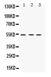
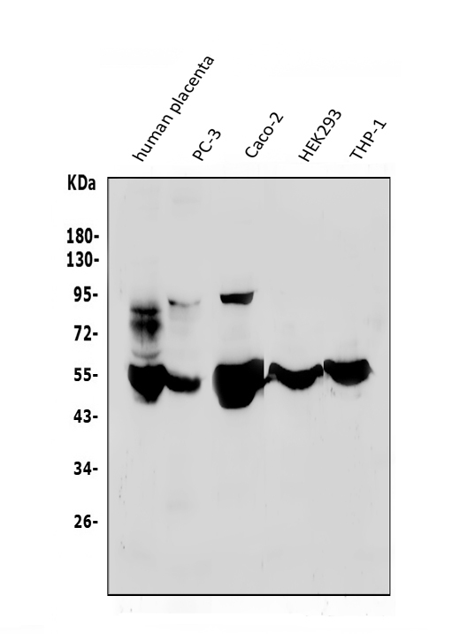
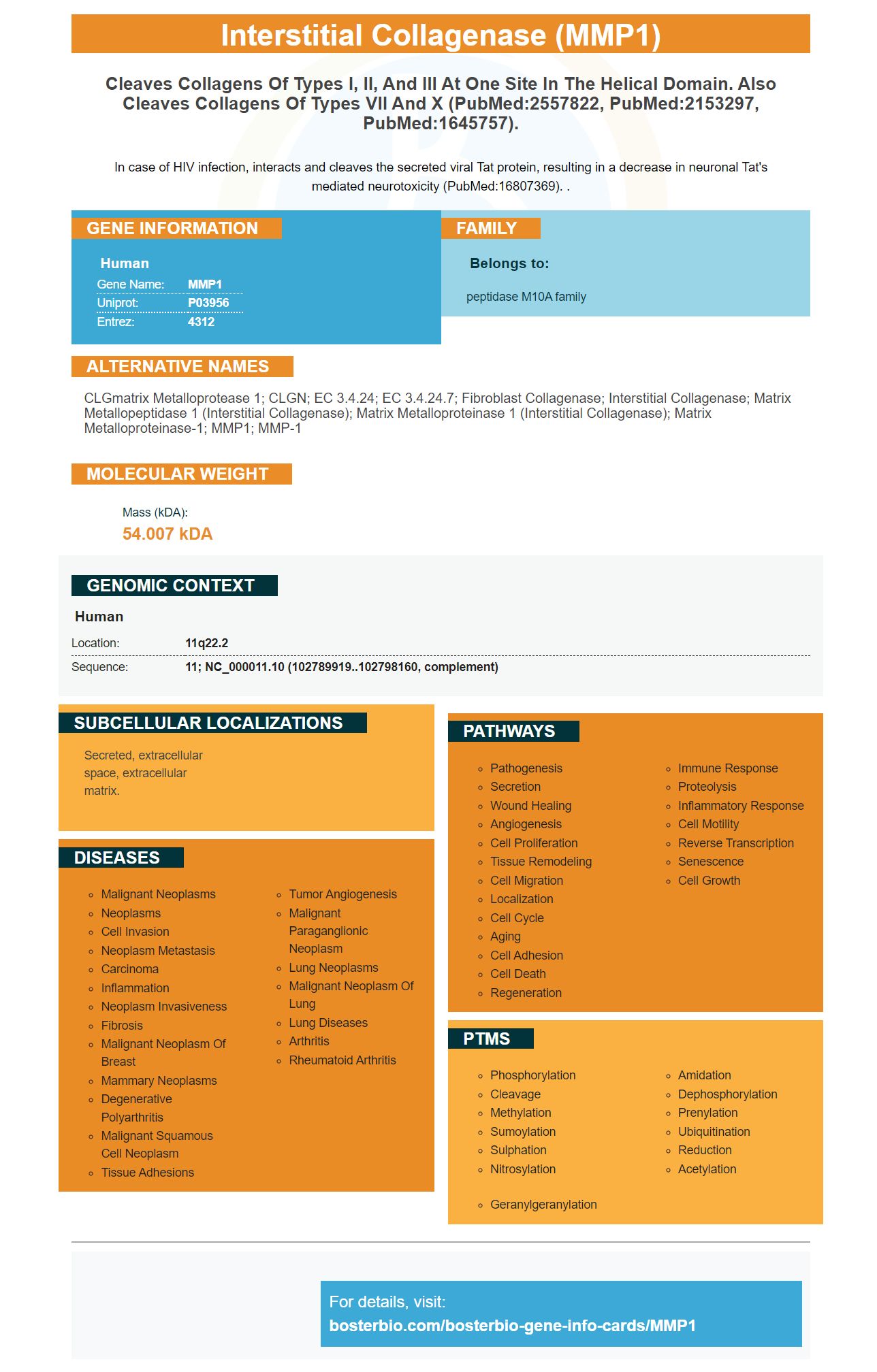
Facts about Interstitial collagenase.
In case of HIV infection, interacts and cleaves the secreted viral Tat protein, resulting in a decrease in neuronal Tat's mediated neurotoxicity (PubMed:16807369). .
Human | |
---|---|
Gene Name: | MMP1 |
Uniprot: | P03956 |
Entrez: | 4312 |
Belongs to: |
---|
peptidase M10A family |
CLGmatrix metalloprotease 1; CLGN; EC 3.4.24; EC 3.4.24.7; Fibroblast collagenase; interstitial collagenase; matrix metallopeptidase 1 (interstitial collagenase); matrix metalloproteinase 1 (interstitial collagenase); Matrix metalloproteinase-1; MMP1; MMP-1
Mass (kDA):
54.007 kDA
Human | |
---|---|
Location: | 11q22.2 |
Sequence: | 11; NC_000011.10 (102789919..102798160, complement) |
Secreted, extracellular space, extracellular matrix.
If you are looking for information about the Bradford assay and the MMP1/TIMP1 complex, take a look at this article. This article will provide an overview of the Bradford assay and what you can expect from it. You will also learn how to use the MMP1/TIMP1 marker. Continue reading to learn about the best uses for this biomarker. Here are some examples.
The MMP1 protein, which is widely studied, plays an important role in both tissue remodeling and resorption. The MMP-TIMP balance is disturbed in various pathological conditions. This can cause MMP levels to become abnormal. The identification of MMPs in biological samples is a key element in diagnosing and the development of new treatments. In this article, we'll discuss the different methods used to detect MMPs.
Light microscopy is the first method for detecting MMP activity. In light microscopy, MMP activity is represented by white spots on black backgrounds. The second method involves switching between catalytic zinc and catalytic zinc to form an intermediate active enzyme. This enzyme is degraded further upon activation by autolytic cleavage as well as the activity of other proteases. This results in a reduction of the MMP pro-domain with an 8-10kD molecular mass. The MMP family is also activated by other proteinases such as agglutinin or tyrosinephosphatase.
Reverse zymography also detects the MMP1 protein. This technique employs the same technique as MMPs, but is more sensitive. TIMPs have been linked to various pathological conditions, including cancer infections, infectious diseases, and ageing. TIMPs have different roles to play in the process of tissue remodeling and the balance influences the normal physiological processes. It is crucial to understand that TIMPs are not extensively studied and may have clinical value as an alternative to MMP inhibitors.
MMP1 is a vital enzyme protein that can be used to diagnose various diseases such as cancer. In the past, researchers used Tissue immunostaining as well as serum levels of gelatinases to identify cancers of the esophageal region. The results of these studies show that MMP1 is overexpressed in many types of cancer which includes esophageal carcinoma. However the enzyme is not commonly used to detect cancer.
The detection of ESCC through the use of MMP1 from plasma was effective. However, this study was a cross-sectional study and it was not able to establish a causal link between MMP1 expression and presence of ESCC. This is why researchers are still examining ways to utilize MMP1 to detect ESCC in a clinical setting. Multiple studies have shown that MMP1 expression can increase detection of ESCC.
MMP-1 is associated with fibrosis. Its level is typically lower than in the control group. Leroy et al. found that MMP-1 levels in serum are ininverse relation to the severity of liver fibrosis. The MMP-1 marker demonstrated an opposite correlation to the METAVIR scores for fibrosis. Additionally, patients with F3 and F4 cirrhosis had lower levels of MMP-1 compared to those who had a fibrosis grade of F1 or F2.
Multifunctional enzymes, MMPs, play a variety of roles in a myriad of ailments. They are known to impact inflammation, wound healing, and tumorigenesis. An increase in MMP1 expression is linked to inflammation, cancer, as well as chronic degenerative diseases. Most of the collagenases in the body are present in macrophages and epithelial cells. Ras/Raf may activate the MMP1 gene.
The MMP1/TIMP1-complex is a highly versatile enzyme that plays an important role in normal tissue remodeling as well as tissue loss in pathological conditions. These enzymes are important for the diagnosis and developing new treatments. In biological samples, there exist a variety of types of MMPs. These proteins can be identified with the Boster Bio enzyme test.
The Boster Bio MMP1/TIMP1 Complex contains the enzymes MMP-2 and TIMP-1. They are detected in the conditioned media. The MMP-TIMP1 complex is separated by SDS and then moves through the gel in accordance with their respective molecular mass. The Coomassie stained gel shows three separate sections: one of which is devoid of MMPs nor TIMPs, the third one with both.
Gel substrate zymography is used to determine the activity of the enzyme. MMP-2 activity was highest at 5 NIH U/mL thrombinlevels, which is 3.2 +/0.5 fold higher than the control group. The latent MMP-2 variant was observed in the EC without stimulation. The enzyme was found to possess molecular masses that ranges from 87 to 100 kD. It was also discovered that 1,10-phenanthroline was able to remove the gelatinolytic bands from ECs when added to the buffer for substrates.
The role of thrombin in activation of MMPs is a major focus of the study. Understanding how thrombin affects MMP activity is crucial to the development of new drugs to target angiogenesis and the growth of tumors. The team of researchers will study the activities of MMPs in various tissues and investigate the effects of thrombin upon the protease. This data will facilitate the creation of new inhibitors of thrombin that target these enzymes once it is available.
The Boster Bio Bradford assay is among several protein tests available on the market today. This method employs a standard bovine serum albumin to determine the concentration of protein in a sample. In some cases other protein standards could be used, as well. Samples are deionized water prior to assaying. Bio-Rad's Bradford reagent is diluted 2.5 times and then added to three wells.
The Bradford assay measures the concentration of proteins in solutions as low as 1 mg to 20 mg. This method is extremely sensitive because the reagent is stored in phosphoric acid for up to two weeks before it begins to degrade. The dye also generates a graph that can be read at 595nm wavelength. The Bradford test is fast and easy. The slope of the line determines the protein content.
The Bradford assay is based off Coomassie G-250, which is an infrequently used in protein gels. This reagent resists acid, and it is able to bind protein in equilibrium pH ranges between 0.7 and 1.0. Its sensitivity is comparable to the Bio-Rad DC Protein Assay however, it is more sensitive. In this way, it's the most accurate method to determine the amount of protein.
To determine the activity of MMP1 protein SDS-PAGE is a popular method to observe changes in the levels of proteins in the blood. This method uses protein markers that include all subunits as well as proteins. The proteins are lyophilized, and then reconstituted in deionized waters. The protein markers create bands that are well-defined and have the same intensity.
The process of SDS PAGE involves the polymerization of the acrylamide when it is in contact with a substrate. Other substrates are also able to determine MMP activity in addition to gelatin. The special polyacrylamide gel is then electrophoresed at 37°C in an acidic buffer composed of Zn2+ and Ca2+. During electrophoresis with SDS MMPs are activated.
The specificity loop is the shortest within the MMP family, and it ends at Gly261 with an a-helix hC. The catalytic domain is surrounded by the a-helix and hC. SDS can attach multiple MMPs to the same strand, and this method is reliable for identifying specific MMPs. SDS-PAGE can be used to analyze the activity of a specific MMP.
Gelatin zymography has demonstrated an increase in dose-dependent amount of MMP-2 and MMP-9 proteins. MMP-2 and MMP-9 proteins were the most preferred substrates. The results showed that MMP-2 and MMP-9 showed concentration-dependent bands. These results indicate that SDS-PAGE could be used to measure MMP activity within cells. The proteins could be utilized in the near future to create therapeutic applications.
The mROC program is a computer program which determines the sensitivity and the specificity of markers, determines the optimal combination and produces the highest results. Two or three markers are usually the most effective combination. Even the most sensitive and precise indicators may not be the most effective. MMP2 by itself will not provide a satisfactory level of sensitivity. However, MMP9 can increase the specificity and sensitivity up to 95%.
MMP1 plays a role in the breakdown of the extracellular matrices. Extracellular matrix plays a significant part in various physiological processes , as well as disease. Particularly, this enzyme breaks down interstitial collagens which are a component of cartilage. It also can contribute to corneal permeability, which can contribute to the growth of cancer. This kind of enzyme has an impact on the process of metastasis and thus determining its expression may help identify tumors early.
A study published in Molecular Pathology Journal (2007) used blood plasma samples taken from 68 patients suffering from TCC in the bladder to assess the MMP1/TIMP1-complex. The mROC program was utilized by the authors to identify the two- and three-marker combinations with the highest correlations between the two markers. The overall diagnostic performance index as well as the area under the ROC curve and the sensitivity and specificity at the 95 percent and 90 limit of detection were used to determine the diagnostic values for single markers.
PMID: 2167156 by Templeton N.S., et al. Cloning and characterization of human tumor cell interstitial collagenase.
PMID: 3030290 by Whitham S.E., et al. Comparison of human stromelysin and collagenase by cloning and sequence analysis.
*Showing only the more recent 20. More publications can be found for each product on its corresponding product page