This website uses cookies to ensure you get the best experience on our website.
- Table of Contents
1 Citations 6 Q&As
Facts about Mitofusin-1.
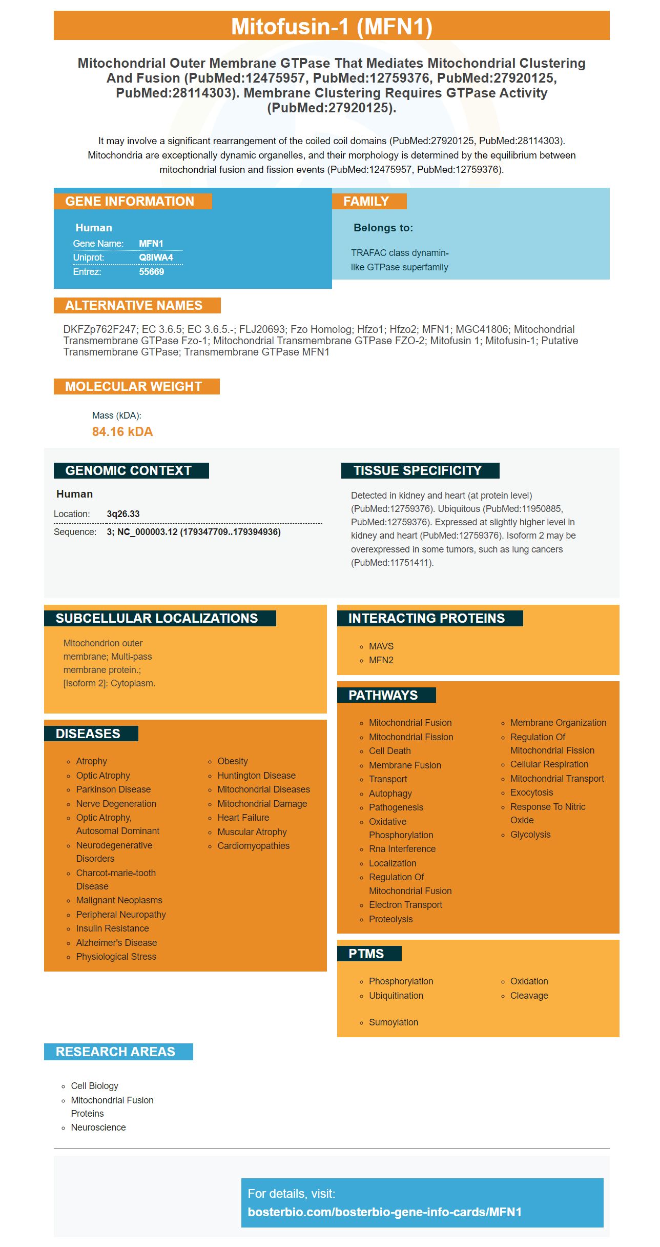
It may involve a significant rearrangement of the coiled coil domains (PubMed:27920125, PubMed:28114303). Mitochondria are exceptionally dynamic organelles, and their morphology is determined by the equilibrium between mitochondrial fusion and fission events (PubMed:12475957, PubMed:12759376).
Human | |
---|---|
Gene Name: | MFN1 |
Uniprot: | Q8IWA4 |
Entrez: | 55669 |
Belongs to: |
---|
TRAFAC class dynamin-like GTPase superfamily |
DKFZp762F247; EC 3.6.5; EC 3.6.5.-; FLJ20693; Fzo homolog; hfzo1; hfzo2; MFN1; MGC41806; mitochondrial transmembrane GTPase Fzo-1; mitochondrial transmembrane GTPase FZO-2; Mitofusin 1; mitofusin-1; putative transmembrane GTPase; Transmembrane GTPase MFN1
Mass (kDA):
84.16 kDA
Human | |
---|---|
Location: | 3q26.33 |
Sequence: | 3; NC_000003.12 (179347709..179394936) |
Detected in kidney and heart (at protein level) (PubMed:12759376). Ubiquitous (PubMed:11950885, PubMed:12759376). Expressed at slightly higher level in kidney and heart (PubMed:12759376). Isoform 2 may be overexpressed in some tumors, such as lung cancers (PubMed:11751411).
Mitochondrion outer membrane; Multi-pass membrane protein.; [Isoform 2]: Cytoplasm.
If you're considering purchasing the MFN1 marker, you should first be aware of the MFN1 protein's function. It interacts with Pink to trigger autophagy within cells. It also modulates the rate of proliferation and migration of LAD cells at high glucose levels. MFN1 also plays a role in the regulation of Fis1 and Parkin expression. Boster Bio has more information about the MFN1 marker.
MFN1 is a crucial member of the MAMP (Mitochondrial Autophagy MAP) family. It plays an important role in mitochondrial autophagy , by interfacing with Pink and regulating the membrane potential of the organelle. MFN1 has been implicated with the pathological effects of glucose EMT (LAD) It also has multiple roles in the ER.
MFN1 can be induced by high glucose and is a latent mediator of a variety of biological processes involved in cancer. We're not certain of the mechanisms that regulate the expression of this protein. This study looked into Mitofusin1's role in high glucose-induced EMT (LAD cells) in this study. It has identified a molecular mechanism that regulates the function of MFN1 in this process.
In NAFLD the cardiac mitochondrial function decreases when there is MFN1. However, exercise increases the autophagic flow in the heart. The improved function of the heart is associated with improved mitochondrial bioenergetics. As a result, exercise can aid in reversing heart failure. But, can exercise be used to stop NAFLD? Possibly. Exercise is the solution.
The antigen is derived from rabbits and mice, and may be monoclonal or polyclonal in nature. This antibody can recognize both MFN1 as well as MFN2 in different animal specimens. However, there is a possibility of significant rearrangements in the coil domains. The mitochondria are extremely dynamic organelles. The balance between fission and fusion events regulates how the organelle performs its functions.
The loss of MFN1/MFN2 in mice results in an increase in autophagic flux. However, overexpression of MFN2 in mice has prevented mitophagy and peripheral nerve damage induces both mitophagy and apoptosis. Although the relationship between MFN1/Mfn2 with cancer isn't clear, anti-cancer drugs that inhibit mitochondrial function also have an impact on mitophagy.
MFN2 and Mfn1 were demonstrated to have a connection to various cancer types. Both are associated with liver disease progression. The absence of MFN1 blocks the activity of the MAPK-ERK-CREB signaling pathway. The drug that targets MFN2 could be beneficial to cancer. Even though both proteins are implicated in the development of liver disease, MFN2's anti-cancer benefits could be beneficial to patients with various forms of cancer.
The dysfunction of autophagy caused by glucose has been connected to A549 cells (a kind of cancerous cell). Autophagy enhances EMT and increases migration, infiltration, and ultimately leads to LAD. Additional mitochondrial proteins are needed to aid in autophagy. These proteins could also serve as molecular scaffolds for the regulation of LAD.
High glucose was added to A549 cells to enhance the MFN1/Pink interactions. We used the STRING database to discover proteins that interact with MFN1. After glucose exposure We then examined the expression of Pink and Parkin in A549 cells. We also used the Western blot assay to determine whether Boster Bio MFN1 is a marker that interacts with Pink and triggers autophagy within A549 cells.
Higher levels of lipids can be linked to autophagy inhibition. A large intake of lipids could cause liver dysfunction. The accumulation of fatty acids also causes ER stress in the hepatocytes. Moreover, MFN2 is thought to serve as a connector between mitochondria and ER. It helps transfer phosphatidylserine from ER and mitochondria.
The MFN1 interaction with Pink is the result of a mRFP/GFP-LC3 fusion protein. The cellular localization patterns of the mRFP/GFP-LC3 fusion proteins in A549 cells have been revealed. Further research will establish if the MFN1 marker is involved in the inhibition of autophagy. This interaction can cause cancer by blocking autophagy.
In addition to interfering with autophagy, MFN1 may also inhibit the growth of cancer cells. The loss of MFN1 has been related to decreased autophagosome creation and the accumulation of damaged mitochondria. The authors found that the loss of MFN1/Mfn2 inhibits autophagy. In mouse cardiomyocytes, mitophagy was affected by pink and LATS2 overexpression.
MFN1 is not just linked to the mRFN1–PINK gene to regulate apoptosis. When exposed to high glucose levels, A549 cells showed an increase in migration and invasion. The inhibition of MFN1 slowed the rate of apoptosis, and reduced levels of MFN1 diminished cell growth. MFN1 siRNA inhibited autophagy however, it decreased the levels of N-cadherin and Snail, two proteins that negatively impact autophagy.
Increasing glucose levels within the body may encourage the migratory behaviour of tumor cells. Tumor cells are constantly migration-oriented, however, hyperglycemia may also promote invasion. These changes in migratory activity could be related to changes in the tumor-specific MMPs or uPA. Inflammatory phenotypes of tumors are marked by an increase in invasion.
EMT is accompanied induction by p38 MAP kinase. Eosinophils are controlled by MAP kinases to regulate the rapid matrix metaloproteinase-9 (MMP) release. Invasive cytotrophoblasts also contain p38 MAPkinase which regulates expression of adhesion molecules. Chapman HA coordinates cell adhesion, and cell migration. Furthermore, Waltz DA promotes cell mobility by facilitating the interaction between the urokinase receptor as well as vitronectin.
In addition, MFN1 modulates high glucose-induced cell apoptosis by dephosphorylating serine 46. It is also well-known that it triggers degrading of HIPK2 proteins. This study highlights the important role of this resourceful oncosuppressor in tumor cells. It is possible that p53 could stop the progression of prostate cancer.
MFN1 also inhibits mitochondrial fission which is an aspect that regulates the cell death rate of human cancers. The effects of high glucose on LAD cell lines can reduce NFKB or TP53, increasing self-phagy. MFN1 could be a new therapeutic strategy to treat human cancer.
It is not clear what MFN1's role is in GDM. The role of MFN1 during the development and progress of the disease has been linked with its association with inflammation and hyperglycemia. This is why this protein has been implicated in the development of preeclampsia an inflammation-related condition. MFN1 is also involved in the pathophysiology of preeclampsia and MR in the walls of vascular vessels.
The Bcl-2 protein family regulates cell migration, invasion, and metastasis. It also blocks the degradation of matrix enzymes and transcription factors. They are abundantly present in the lymph nodes, liver and the lung of patients with breast cancer. These proteins also inhibit cell migration, invasion and growth of osteosarcoma as well as breast cells. Their roles may differ among patients.
MFN1 inhibition results are reprogramming and increase in glucose metabolism in tumours. This non-invasive test of glucose uptake has shown numerous clinical applications for treatment and diagnostics of cancer. MFN1's unique role in creating EMT can be attributed to its interaction with Pink which induces autophagy. This abnormal autophagy is the cause for the pathological EMT in A549 cell lines.
The inhibition of MFN1 also reduces the expression of Pink which is a protein found in the outer membrane of most cells. It is vital to the mitochondrial membrane potential and aids in mitochondrial energy metabolism in addition to its cancer-promoting properties. Pink stimulates autophagy receptors via the LC3 interfacing region motif. This triggers autophagy, and forms a binding bond with the LC3B II version. The interaction between MFN1 and Pink also improves autophagy.
High glucose increased the migration and invasion of A549 cell lines. The effects of high glucose were slowed by transfection of MFN1. It also inhibited N-cadherin expression in A549 cells. It is possible that MFN1 plays a role in the pathogenesis or development of the LAD. The results also support the role played by MFN1 in cellular migration and invasion.
In addition to peptide kinase and Fis1 inhibitors, Boster Bio also offers ELISA kits for measuring these proteins. They have been thoroughly tested and are trusted by the research community. Moreover, they have been validated on Western Blotting, Immunohistochemistry, and ELISA. Boster Bio's MFN1 inhibitors are a good option for anyone who is interested in research on Parkinson's disease.
In addition to the effects of MFN1 inhibitors on the NF-B pathway, Parkin inhibits mtDNA depletion. In addition, it stimulates autophagy in mitochondria. The inhibitors also inhibit Parkin and Fis1 expression. The inhibitors are very efficient in preventing PRRSV-induced depletion of mtDNA in human liver cancer cells.
The research also shows that Drp1 interferes with the mitochondria's function. Drp1 also influences neurotransmission, mitochondrial fusion, and the axon. Parkin and Fis1 are proteins essential for neurodegeneration and mitophagy. Inhibiting Drp1 reduces both Parkin as well as Fis1 expression in neurons.
PMID: 11181170 by Santel A., et al. Control of mitochondrial morphology by a human mitofusin.
PMID: 9230308 by Hales K.G., et al. Developmentally regulated mitochondrial fusion mediated by a conserved, novel, predicted GTPase.
*More publications can be found for each product on its corresponding product page