This website uses cookies to ensure you get the best experience on our website.
- Table of Contents
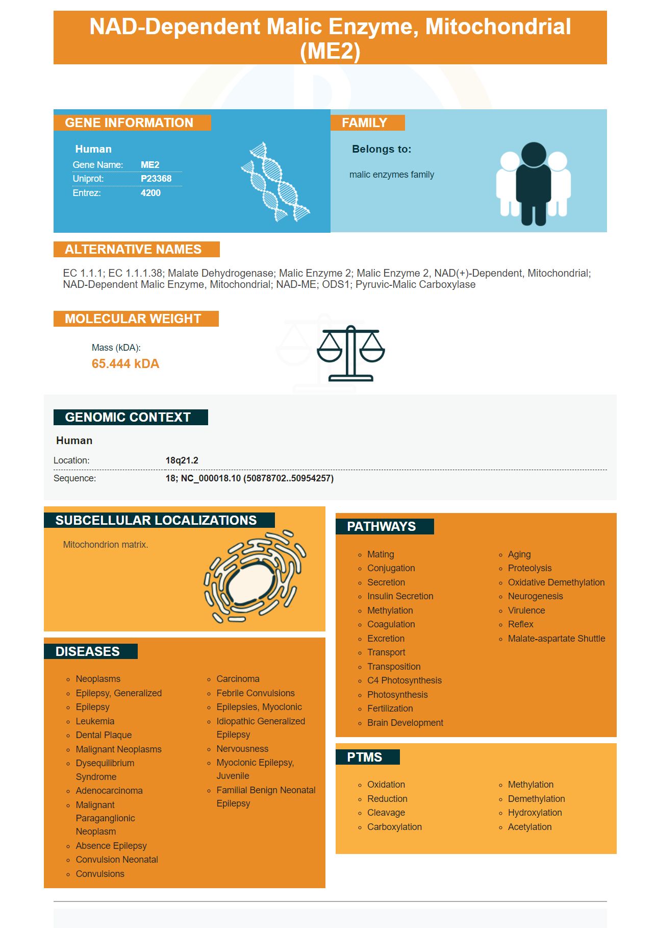
Facts about NAD-dependent malic enzyme, mitochondrial.

| Human | |
|---|---|
| Gene Name: | ME2 |
| Uniprot: | P23368 |
| Entrez: | 4200 |

| Belongs to: |
|---|
| malic enzymes family |

EC 1.1.1; EC 1.1.1.38; malate dehydrogenase; Malic enzyme 2; malic enzyme 2, NAD(+)-dependent, mitochondrial; NAD-dependent malic enzyme, mitochondrial; NAD-ME; ODS1; pyruvic-malic carboxylase
Mass (kDA):
65.444 kDA

| Human | |
|---|---|
| Location: | 18q21.2 |
| Sequence: | 18; NC_000018.10 (50878702..50954257) |
Mitochondrion matrix.


Described in this article are the origins, interrelationships, and clinical applications of the ME2 Marker and Steven Boster. For more information on the ME2 Marker, read Boster Bio The Best Uses For The ME2 Marker. Steven Boster is a scientist who invented the ME2 Marker. He developed the first primary antibody in 1993. Since since then, he's created hundreds of them. By the late nineties, Boster was the most powerful catalog antibody manufacturer in China. Boster also developed a proprietary ELISA platform, PicoKine(tm), and employed his own trade secrets to deliver high-sensitivity ELISA kits.
There are numerous ways to analyze the ME2 marker. Knockdown experiments revealed that ME2 knockdown inhibits invasion in a transwell-BD matrigel test. These results indicate that ME2 plays a significant role in cell growth, migration, and invasion. Furthermore, knockdown studies showed that ME2 inhibits LN229 cell migration. These results suggest that ME2 is a crucial component of cancer cell proliferation and migration.
Different types of gliomas have ME2 expression in different types of gliomas. In addition to the detection of its expression in various tumors, ME2 is also correlated with other mesenchymal markers found in tumors called gliomas. ME2 expression was identified using Western Blotting and real time PCR. These tests also reveal the presence of PN markers and the MES marker. Physicians can use the ME2 marker to diagnose GBM and other cancers.
In addition to its lipid-binding capabilities, ME2 has been shown to enhance the process of lipogenesis in glioma cells. It inhibits the production reactive oxygen species and AMPK phosphorylation. It also promotes ACSS2 lipogenesis. increases SREBP-1 nuclear location. It is not clear whether ME2 is involved with the development of GBM. However it is a unique classification marker that could help doctors find a new treatment.
ME2 increase in mice led to a decrease in expression of epithelial markers and PN. In contrast, ME2 knockdown produced opposite results. ME2 stimulates the production of PMT and blocks ROS in mitochondria. Western Blots confirmed the presence of E, N, cadherin and vimentin. YKL-40 and OLIG were also confirmed. A fluorescent probe also revealed ROS in mitochondria. The color green is a sign of oxidized components in lipids, while the red color indicates nonoxidized components.
Boster is the best option if you are looking for a high-affinity primary anti-ME2 antibody. With a 25-year track record of quality and reliability, Boster antibodies are cited frequently in scientific journals and are trusted by researchers all over the world. In addition to their high affinity, Boster antibodies have been validated for use in Western Blotting, Immunohistochemistry, and ELISA.
The ME2 marker is found in oral squamous cell carcinoma (OSCC) tissue in high amounts. Immunohistochemical staining allows for the detection of the expression of the whole slide. It can be detected in primary and secondary oral squamous cell carcinoma tissues. OSCC survival is related to the ME2 expression level. Clinical applications of the ME2 marker have been created to determine the rate of progression of the disease.
The expression of ME2 in cancer tumors has been shown to affect the effectiveness of therapy. In addition the expression of ME2 has been found to be associated with clinicopathological significance in OSCC. Studies of human OSCC tissues demonstrated that ME2 expression was related to mitochondrial respiration. This may be a factor in the pathogenesis of cancer. The high levels of ME2 in cancer tissues may result from an abnormal mitochondrial respiratory process, resulting in increased the severity of the disease and size of the tumor. A high level of ROS may also contribute to the growth of tumors.
Researchers found that HNSCC had a higher level ME2 than the average person, which was linked to a lower chance of survival. However, this association has only been confirmed in a cohort of Chinese people. Further studies are required to confirm the findings. ME2 expression may be helpful in predicting survival rates for cancer. The study was successful because the ME2 expression was detected in patients. The researchers confirmed that ME2 expression is not dependent on smoking and alcohol.
The ME2 marker has five potential applications for the treatment of cancer patients. They could be used for screening, diagnosis, monitoring therapy and detecting the possibility of relapse. Their sensitivity and specificity will determine their value in these scenarios. Researchers have also discovered that the ME2 marker can be used to screen for occult carcinoma. However, these tumor markers have shown only limited utility in early cancer diagnosis. This suggests that ME2 is only useful for cancers that can be detected by other methods.
The ME2 marker, which is located on chromosome 6, is implicated in a variety neurological diseases. Incredibly, it has been linked to myoclonic seizures as well as JAE and EGTCS. Greenberg et al. They identified the ME2 locus as a susceptibility marker for these disorders. This genetic marker is associated to two epilepsy types: petit mal, and myoclonus atypical.
Alongside being crucial for maintaining homeostasis, ME2 is also vital for promoting the PMT process in GBM cells. The ME2 marker inhibits AMPK activity and promotes SREBP-1 nucleolocalization. Additionally, ME2 is associated with PMT which is a reprogramming process of lipogenesis. Future studies that seek to discover the role of ME2 in GBM could result in a new treatment.
Molecular analysis of human glioma cell lines revealed that ME2 expression ME2 was significantly correlated with MET and PN markers. ME2 expression was determined using western blotting and RNA-seq. Expression of ME2 was found to be greater in the U87MG and LN229 cells than in other human glioma cell lines. While ME2 is found in a larger range of human glioma cells, its expression was low in the melanoma cell line.
PMID: 1993674 by Loeber G., et al. Human NAD(+)-dependent mitochondrial malic enzyme. cDNA cloning, primary structure, and expression in Escherichia coli.
PMID: 11401430 by Yanaihara N., et al. Physical and transcriptional map of a 311-kb segment of chromosome 18q21, a candidate lung tumor suppressor locus.


