This website uses cookies to ensure you get the best experience on our website.
- Table of Contents
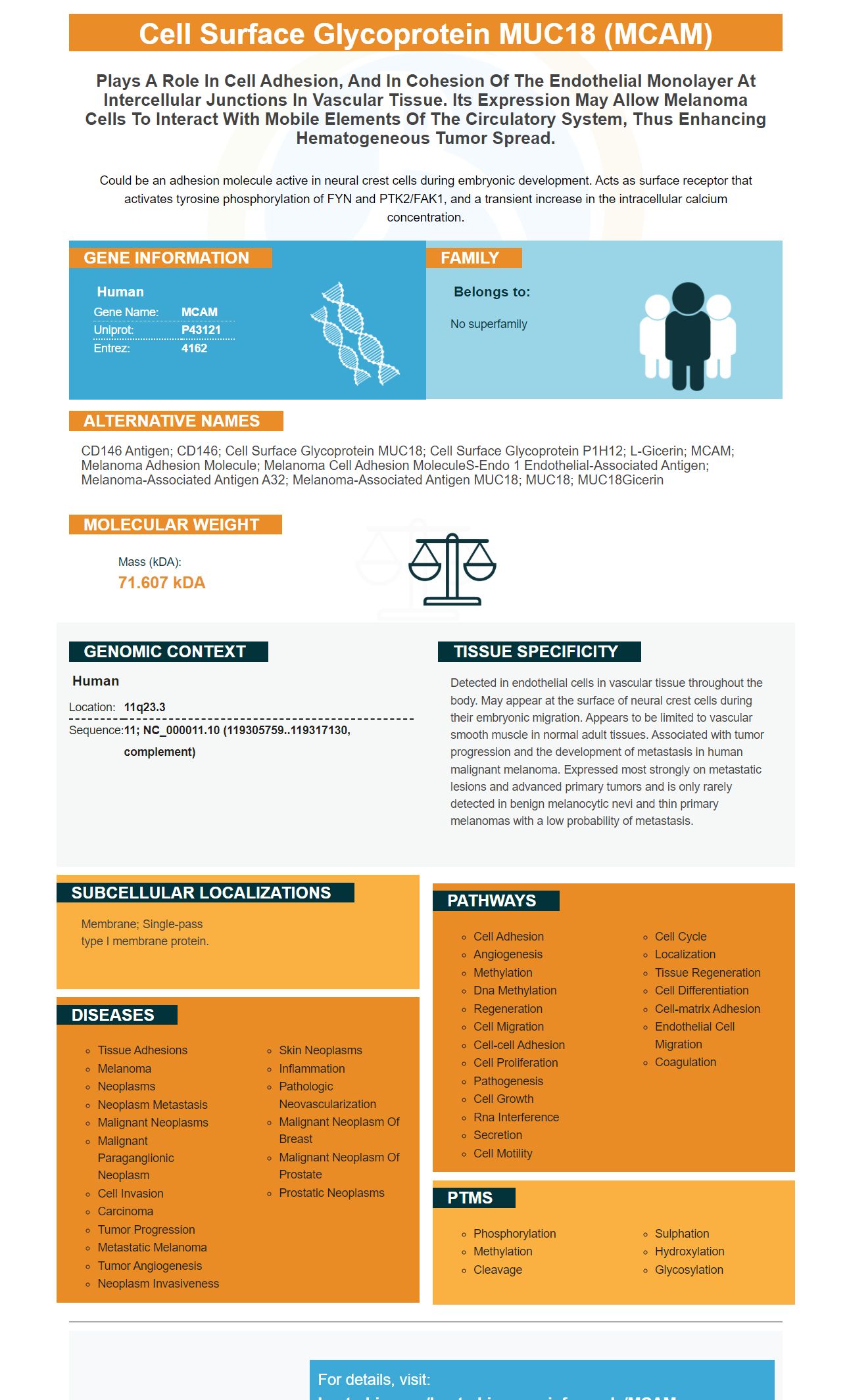
Facts about Cell surface glycoprotein MUC18.
Could be an adhesion molecule active in neural crest cells during embryonic development. Acts as surface receptor that activates tyrosine phosphorylation of FYN and PTK2/FAK1, and a transient increase in the intracellular calcium concentration.
Human | |
---|---|
Gene Name: | MCAM |
Uniprot: | P43121 |
Entrez: | 4162 |
Belongs to: |
---|
No superfamily |
CD146 antigen; CD146; cell surface glycoprotein MUC18; Cell surface glycoprotein P1H12; L-Gicerin; MCAM; melanoma adhesion molecule; melanoma cell adhesion moleculeS-endo 1 endothelial-associated antigen; Melanoma-associated antigen A32; Melanoma-associated antigen MUC18; MUC18; MUC18Gicerin
Mass (kDA):
71.607 kDA
Human | |
---|---|
Location: | 11q23.3 |
Sequence: | 11; NC_000011.10 (119305759..119317130, complement) |
Detected in endothelial cells in vascular tissue throughout the body. May appear at the surface of neural crest cells during their embryonic migration. Appears to be limited to vascular smooth muscle in normal adult tissues. Associated with tumor progression and the development of metastasis in human malignant melanoma. Expressed most strongly on metastatic lesions and advanced primary tumors and is only rarely detected in benign melanocytic nevi and thin primary melanomas with a low probability of metastasis.
Membrane; Single-pass type I membrane protein.
This article will discuss Boster Bio's Anti-Ep-CAM / CD326 (Epithelial Marker) and Boster Bio Anti-NCAM1 Antibody Picoband. Both of these products target MCAM and are highly specific. This article will also provide an overview of the best uses of Boster Bio's Anti-NCAM1 Antibody Monoclonal.
Boster Bio Anti-NCAM1 Antibodies are monoclonal antibodies that react with the MCAM marker on human, mouse, and rat cells. Each antibody is derived from E. coli and is tested on immunofluorescence and WB. This product comes with a blocking peptide at 1.0mg/l. If you plan to use large amounts of samples, you should consider buying a multichannel pipette.
The G8.8 monoclonal antibody recognizes the 40 kDa EpCAM protein. Also known as EPCAM1, EpCAM2, LAIR-1, or TACSTD1, EpCAM has been identified on a wide variety of human cancers. Its role in carcinogenesis has not been fully explored, but recent discoveries at the First International Symposium on EpCAM Biology and Clinical Application are changing our understanding of this vital protein.
This single-pass type I glycoprotein is expressed on the membranes of most epithelial cells. It colocalizes with CD326, CLDN7, and integrin. Although EpCAM and TROP-2 share nearly 50% sequence similarity, their functions are vastly different. As such, it is best to avoid referring to these proteins as EpCAM-1 and EpCAM-2.
Using this antibody, we detected the EpCAM protein in PC-3 cells. These cells have been sown on 96-well poly-L-lysine microplates. We then used these cells as antigens in an ELISA. We selected a hybridoma clone that showed a strong reaction against EpCAM.
The CP region of EpCAM is responsible for interacting with human cells. The present inventors intensively examined the iCeap described in Non-Patent Document 1 to develop a better product. This product contains two antibodies, HEA-125 and EBA-1, and one inhibits the binding of the second antibody.
This antibody is highly specific for epithelial tissue. Using it for this purpose requires less than 0.5 ug per test. A test is defined as the amount of antibody needed to stain 100 uL of cells. The number of cells to stain should be determined empirically and can vary from 105 to 108 cells. This antibody is filtered through 0.2 um after manufacture.
This antibody recognizes epithelial cells and their circulatknownu tnrmirs. The kit also detects tumor cells circulatknowin human blood, such as hybridomas. Its accession number is NITE BP-1449. When used with a specific antibody, Boster Bio Anti-Ep-CAM / CD326 (Epithelial Marker) is a highly sensitive and actnrate test.
This antibody targets EpCAM, a membrane protein involved in oncogenic signalling. Its interaction with other proteins may provide a therapeutic window for EpCAM-directed immunotherapies. EpCAM inhibits other proteins in tumour cells, which might open a therapeutic window. Future studies must better understand the relationship between EpCAM signalling and the proliferation of tumour cells.
In a recent study, a human EpCAM-specific IgG1 antibody called Proxinium exhibited tumour-specific immune responses in a pivotal phase II/III clinical trial. In a previous study, this antibody inhibited tumour growth and induced complete response in 25 percent of patients. Additionally, patients treated with EpCAM-specific immunotherapy tended to live 301 days longer than patients in the historic control group.
Monoclonal antibodies are produced by a process called cell-fusion. Kohler and Milstein developed this method and merged spleen cells from animals with EpCAM-reactive myeloma to form a hybridoma group. From there, a monoclonal antibody can be selected from the hybridoma culture.
The company, Boster Bio, is an innovator of high-sensitivity and high-specificity antibodies. Over the past two and a half decades, Boster Bio has honed its technology and techniques to deliver high-quality products that meet the needs of researchers and scientists worldwide. Boster Bio antibodies are characterized by high affinity and have been validated for WB, ELISA, and Flow Cytometry applications.
The company, founded in 1993, makes antibodies and supplies ELISA kits, detection kits, and other products that are compatible with their antibodies. Boster Bio's Anti-NCAM1 Antibody is available in a variety of sizes and is compatible with Human, Mouse, and Rat samples. Boster Bio also offers custom services and BeNeLux deliveries. However, not all of their products are suitable for research purposes.
PMID: 2602381 by Lehmann J.M., et al. MUC18, a marker of tumor progression in human melanoma, shows sequence similarity to the neural cell adhesion molecules of the immunoglobulin superfamily.
PMID: 8378324 by Sers C., et al. Genomic organization of the melanoma-associated glycoprotein MUC18: implications for the evolution of the immunoglobulin domains.