This website uses cookies to ensure you get the best experience on our website.
- Table of Contents
1 Citations
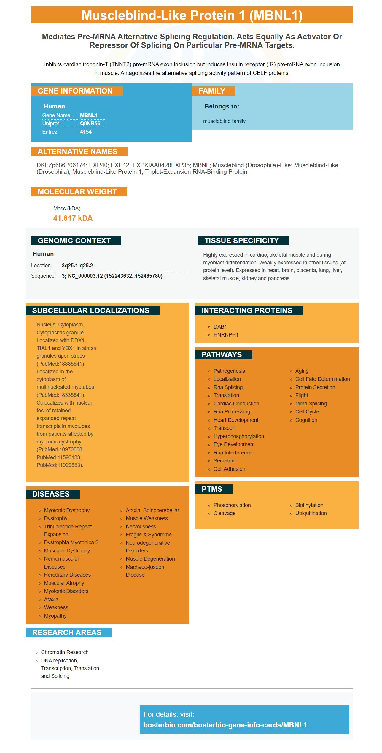
Facts about Muscleblind-like protein 1.
Inhibits cardiac troponin-T (TNNT2) pre-mRNA exon inclusion but induces insulin receptor (IR) pre-mRNA exon inclusion in muscle. Antagonizes the alternative splicing activity pattern of CELF proteins.
Human | |
---|---|
Gene Name: | MBNL1 |
Uniprot: | Q9NR56 |
Entrez: | 4154 |
Belongs to: |
---|
muscleblind family |
DKFZp686P06174; EXP40; EXP42; EXPKIAA0428EXP35; MBNL; muscleblind (Drosophila)-like; muscleblind-like (Drosophila); muscleblind-like protein 1; Triplet-expansion RNA-binding protein
Mass (kDA):
41.817 kDA
Human | |
---|---|
Location: | 3q25.1-q25.2 |
Sequence: | 3; NC_000003.12 (152243632..152465780) |
Highly expressed in cardiac, skeletal muscle and during myoblast differentiation. Weakly expressed in other tissues (at protein level). Expressed in heart, brain, placenta, lung, liver, skeletal muscle, kidney and pancreas.
Nucleus. Cytoplasm. Cytoplasmic granule. Localized with DDX1, TIAL1 and YBX1 in stress granules upon stress (PubMed:18335541). Localized in the cytoplasm of multinucleated myotubes (PubMed:18335541). Colocalizes with nuclear foci of retained expanded-repeat transcripts in myotubes from patients affected by myotonic dystrophy (PubMed:10970838, PubMed:11590133, PubMed:11929853).
The MBNL1 marker is a good option for researchers who wish to differentiate SP and NSP cells from MSC. It has been validated on multiple platforms, with both known positive and negative samples. It is also extremely specific and has a high affinity to its target. Boster is an innovator in the development of immunoreagents. The company's first-time reviews receive credit for the product. This incentive is extended to all scientists in the world.
Immunoprecipitation of anti-MBNL1 antibody along with control IgG was used to identify the MBNL1 protein. The immunoprecipitation method required the synthesis of siRNA duplexes with the following sequences: 5'-CCUGGCAGGGAUUGUAGA-3'32 and 5'-CCUGGCAUUGAGAGAG-3'32. The sequences of MBNL1Ndel1's reporter antibodies were analysed by PCR using MBNL1's intronic region and FACS analysis.
The MBNL1 marker is highly expressed in VSMC but very little is expressed in VSMC–M. This resulted in an increase in macropinocytosis aswell in the expression of a variety of macrophage biomarkers. Moreover, CD68 and aSMA levels were reduced due to the absence of MBNL1.
MBNL1-labeled antibodies colocalized MBNL2 and MBNL3. They also colocalized with MBNL2 as well as MBNL3. The MBNL1 marker as well as the CAGG10 probe were used to observe the protein in DM2 skin fibroblasts. The images were fixed with acetone-methanol and were visualized as grayscale inverted.
These results demonstrate that the MBNL1 proteins regulates various cell-mediated pathways. In addition to the MBNL1 mAb this antibody that targets the MBNL1 marker is highly specific to the Abi1 protein. It recognizes Ndel1 as well as MBNL1, but is not specifically for Abi1–De10.
The expression of Mbnl1 is regulated during the development of the erythroid terminal. Mbnl1 knockdown causes an erythroid differentiation bloc. Mbnl1 inhibits developmentally Ndel1 skipping of the mRNA exon. In addition, knockdown of Mbnl1 interferes with Ndel1 binding of MBNL1. Mbnl1 is therefore essential for the proliferation of erythroids and differentiation.
Expanded repeat foci in adult muscles cells can sequester the MBNL1 protein. Adult muscle has a higher amount of DMPK expression than the juvenile muscle. MBNL1 and MBNL2 are present in nucleoplasmic regions of myoblast cell culture and in the nucleus of cultured myoblasts of adult humans. Microdensitometry has shown reduced levels of MBNL1 and MBNL2 in DM1 and age-matched controls. In addition the MBNL1 mAb is significantly more sensitive to DM1 nuclei than background controls against MBNL3.
Monospecific anti-Mbnl3 antibodies are used to detect regenerating muscle fibers. They recognize an epitope that is present in the majority of Mbnl3 isoforms but not Mbnl1 or Mbnl2. A study was conducted to discover the alternative splicing patterns of mouse Mbnl3 across a variety of tissues. The cloning of cDNA and the sequencing showed that multiple Mbnl3 proteins that had distinct C-terminal exon 7 were detected in various tissues. Additionally, the mRNA for MBNL3 included exon 7C meaning that the mRNA was able to contain the majority of MBNL2 genes.
IHC-optimized, MBNL1-targeting polyclonals can be used to immunohistochemically stain cells in vivo. In this study, the endometrial tissue from nonpregnant macaques of reproductive age was stained by 3 polyclonal antibodies that were produced against different peptide epitopes in human RXFP1. These antibodies were used to visualize endometrial stromal cells.
It is important to maximize the concentration and time of an antigen prior to using it. A well-designed antibody will have a higher binding affinity to the antigen, while decreasing background signal. To increase the specificity your IHC staining, use the Novus Biologicals protocol. When using this antibody, keep it incubating for at least 4 hours at room temperature. You can also choose to lower concentration of antibody and increase the time it is incubated.
MBNL1 is a protein that exists in the human brain tissue. The polyclonal antibody detects this antigen by binding it. It also binds with p21 protein. This antigen is found in all tissues, but not colon cancer tissue. Utilizing an IHC-optimized polyclonal antibody recognizing the MBNL1 marker can improve the effectiveness of diagnosing of human cancer by approximately 50%.
Antigen-affinity purified antibody reduces background staining and produce more reliable results. These antibodies are affinity purified and can interact with the antigen target. This minimizes background staining and assures reliable results. They are also more sensitive and specific than monoclonal antibodies. The MBNL1 marker is useful in determining the antigen that is of concern.
IHC-optimized polyclonally-directed antibodies against the MBNL1 protein are available from a variety of commercial sources. The quality of the antibodies may vary between commercial sources, and they must be evaluated prior to use. Contact the manufacturer for information on the quality of the antibodies. If you are not comfortable purchasing antibodies from a commercial firm then you can buy them directly from R&D Systems.
Monoclonal antibodies do not appear to be very specific for antigens, therefore they have to be regenerated using preimmune sera from the same rabbits. Additionally, antibodies that were preincubated with excess KLH also eliminated staining of epithelial cells. Thus, monoclonal antibody are extremely useful in the diagnosis of many diseases and conditions.
MBNL1 is a protein with an amino acid length of 388 and the mass of 41.8 kDa. It is a peptide that can be found in both the nuclear and cytoplasmic subcellular locations. This protein is implicated in the autoimmune disease Dystrophia myotonica type 1 and has orthologs in both mice and humans. Numerous ELISA kits are now available as targets for this protein.
RBFOX2-MBNL1-mediated upregulation was confirmed through qPCR analysis and manual end-point PCR. This upregulation occurred beginning at the first week of differentiation and increased significantly between the second and third weeks of differentiation. The researchers concluded that MBNL1 is an effective marker to distinguish SP and NSP cells from MSC. This doesn't prove that RBFOX2-MBNL1 can be used in clinical trials however it does suggest that it could be an effective marker.
We utilized two different primers to conduct the MBNL1 RNAi assay. One of they is labeled using Digoxigenin. The other was labeled using the fluorescent dye. The 2-DDCt method was utilized to determine the expression ratio of the target genes. The results revealed that MBNL1 is a mRNA that binds to both SP and NSP cells.
The MBNL1 gene was discovered to have a direct correlation with expression of a number of genes associated with stemness. It has been proven that cancer stem-like cell types arise from malignant transformation of MSC or Reprogramming of mature cells. MBNL1 could facilitate the tumorigenic differentiation of cells, which is essential in the process of initiation of cancer and metastasis, as well as relapse.
Many switch-like splicing processes which included a modification of the RBFOX2 gene, were detected. In addition, a variety of genes were enriched or inhibited by MBNL1 knockdown. In general, these results suggest the gene can be a useful indicator in determining mesoderm differentiation. It is useful in distinguishing SP and NSP cells from MSC in the event of confirmation in clinical studies.
The research was carried out by the Wolfson Centre for Inherited Muscular Disease (CNRS Institute of Genetique Moleculaire) in Montpellier, France. The same research is also carried out by the Institute of Genetic Medicine, Newcastle University, Newcastle-upon-Tyne (UK). These labs use rat anti-GAPDH monoclonal antibodies at 0.1 mg/mL.
This study was done to determine if the well-studied CSC SP from Ewing sarcoma CADO cell lines differs from the NSP and SP of the same cell line. In addition, we examined this cell line in comparison to bone marrow-derived MSCs, which are the putative cell of source of Ewing Sarcoma.
PMID: 10970838 by Miller J.W., et al. Recruitment of human muscleblind proteins to (CUG)(n) expansions associated with myotonic dystrophy.
PMID: 11590133 by Mankodi A., et al. Muscleblind localizes to nuclear foci of aberrant RNA in myotonic dystrophy types 1 and 2.
*More publications can be found for each product on its corresponding product page