This website uses cookies to ensure you get the best experience on our website.
- Table of Contents
4 Citations
Facts about Mitogen-activated protein kinase 1.
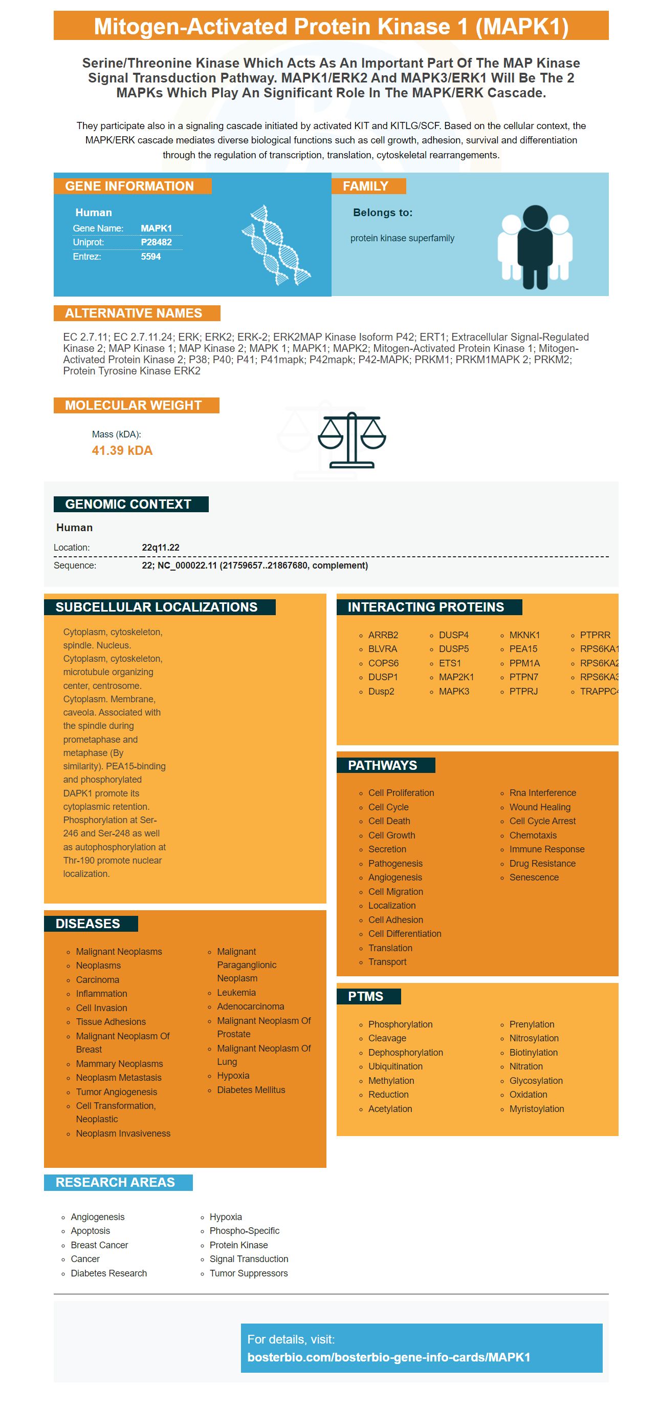
They participate also in a signaling cascade initiated by activated KIT and KITLG/SCF. Based on the cellular context, the MAPK/ERK cascade mediates diverse biological functions such as cell growth, adhesion, survival and differentiation through the regulation of transcription, translation, cytoskeletal rearrangements.
Human | |
---|---|
Gene Name: | MAPK1 |
Uniprot: | P28482 |
Entrez: | 5594 |
Belongs to: |
---|
protein kinase superfamily |
EC 2.7.11; EC 2.7.11.24; ERK; ERK2; ERK-2; ERK2MAP kinase isoform p42; ERT1; Extracellular signal-regulated kinase 2; MAP kinase 1; MAP kinase 2; MAPK 1; MAPK1; MAPK2; mitogen-activated protein kinase 1; Mitogen-activated protein kinase 2; p38; p40; p41; p41mapk; p42mapk; p42-MAPK; PRKM1; PRKM1MAPK 2; PRKM2; protein tyrosine kinase ERK2
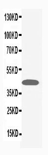
Mass (kDA):
41.39 kDA
Human | |
---|---|
Location: | 22q11.22 |
Sequence: | 22; NC_000022.11 (21759657..21867680, complement) |
Cytoplasm, cytoskeleton, spindle. Nucleus. Cytoplasm, cytoskeleton, microtubule organizing center, centrosome. Cytoplasm. Membrane, caveola. Associated with the spindle during prometaphase and metaphase (By similarity). PEA15-binding and phosphorylated DAPK1 promote its cytoplasmic retention. Phosphorylation at Ser- 246 and Ser-248 as well as autophosphorylation at Thr-190 promote nuclear localization.
Boster Bio is a MAPK1-targeting product that you may be interested in purchasing. This gene targeting product targets MAPK1 across multiple cell types. It can be used in many types of research. If you're looking for a more personalized approach to your research, you should consider a recombinant MAPK1-antibody. The MAPK1 protein targets ERK7 and ERK8 in cells. It also regulates ERK9, ERK12 in cells.
It is known that phosphorylation (of MAPK1IP1L) can trigger a variety of biological assays. These assays use antibodies to MAPK1IP1L. They can be monoclonal, or polyclonal. Boster Bio's MAPK1 antibody reacts with a variety o animal samples. Boster Bio develops its antibodies using rabbit and mouse. The company derived a full-ORF clone resource using the German cDNA consortium.
Orthologs of the human MNK1 protein have also been found in other species. This protein's c-terminal extension is responsible for subcellular location and autoactivation. Its N terminal region promotes its degradation via the Ubiquitin-proteasome pathway. These proteins have been implicated with various disorders and diseases. This discovery led two orthologs to be discovered in the MNK family.
There are many options available to researchers who are looking for a new antibody for studying the human MAPK1 proteins. Boster Bio MAPK1 antibodies are catalog #PA1049. They are highly specific for human MAPK1. It reacts to mouse and rats samples and is stable up to -20 Celsius (-80 F). The product contains five milligrams of BSA, 0.05mg Thimerosal and 0.1mg N3 and can be stored at 20 degrees Celsius for up to one year. This antibody recognizes human MAPK1/3 (171-184aa) for a variety applications.
In biological assays, antibodies detect MAPK1 proteins. The antibodies can be monoclonal or polyclonal and react with MAPK1IP1L in a wide variety of animal samples. BosterBio uses rabbit and mouse as models to develop antibodies to this protein. The German cDNA consortium has provided full-ORF clone resources for the MAPK1 protein.
The MAPKAPK1 mammalian marker plays a crucial role in gene regulatory. A vast array of MAPKKKs are involved in this process. Among the most popular are JNKs, ERK1/2, and p38. However, recent studies have shed some light on the regulation of other groups. Hence, Boster Bio provides educators with educational resources to help them better understand the role of this marker in the body.
The unique cDNA sequence that encodes the MAPK1 genes was derived from a rat's library. The ERK3 cDNA has a C-terminal extension of 178 amino acids and a molecular mass of 100 kDa. Similarly, ERK4 was isolated using the same method, but originally described as a 557-aa protein. Despite their differences in function, they share the same amino acid sequence which indicates that the two proteins may be related.
The MAPK1 gene has been identified as a target for cancer cells. But what are the best uses for this marker? Below are some suggestions on how to use this gene. If you have any questions, please contact Boster Bio. We are here to help. All of our products meet or exceed your specifications. For more information, visit our Boster Bio site.
The MAPK protein family is made up of many proteins that participate in various processes. These proteins include ERK1/2, JNK, and p38MAPK. Each of the proteins has a specific function in the body. ERK1/2 (most commonly activated) and p38MAPK (most frequently disabled) are the two most common kinases. Although ERKs and the p38MAPK have been shown to interact with one other, there is not evidence that they have direct interactions.
The MAPK1 marker is a biomarker for ERK2 and ERK3. ERKs are critical players in cell cycle progression. They regulate cellular growth. The enzymes' enzymatic abilities are dependent on the phosphorylation residues. ERK2 & ERK4 were among the first proteins to be identified as requiring phosphorylation. These proteins are highly relevant to the study of embryogenesis.
The MAPK1 kinase is a key player in regulating cell proliferation and survival. It interacts with the cyclin D1 kinases in order to regulate the G1/S transition. It also interacts SGK to prolong the MAPK cascade, phosphorylating additional members of MAPKAPK's family. RSK, MNK or ERK could be some of these members.
If you're interested in the best uses of the MAPK1 marker, you've come to the right place. ERK1/2 regulates a large number of cellular processes, including neuronal cell differentiation and normal vascular development. ERK1/2 also functions in the cytoskeleton. It is important to understand how this marker works in order to make the most of it.
Many biological assays make use of antibodies that recognize MAPK1IP1L. These antibodies can be monoclonal (or polyclonal) and react with many animal samples. Boster Bio uses rabbit and mouse as model systems to develop its antibodies. These antibodies can be generated using the full ORF clone resource from the German cDNA Consortium.
The MAPK1Marker can be used for a variety biological assays. Boster Bio has created an antibody for human MAPK1IP1L (catalog number PA1049), that reacts with both mouse as well as rat samples. The antibody is stable up to -20degC. It also comes with five milligrams BSA. Thimerosal is also contained in the antibody. It binds to human MAPK1, an antibody that contains a sequence containing 171 amino acid.
For efficient phosphorylation with p38a, the Tyr residue of the ERK1/2 protein is critical. The D domain of MK5 does not need to dock to ERK3/4. Instead, the C-terminal area of the protein contains degenerate sequencings that mediate binding of ERK3/4. Further research is needed to determine the function of this region.
The bosterbio MAPK1 antibody, a monoclonal monoclonal anti-polyclonal antibody, reacts with human MAPK1 proteins, mouse MAPK1 proteins, and rat MAPK1 antibodies. The antibody is stable at temperatures below -20°C for one-year. It contains 5 mg of BSA and 0.05 mg Thimerosal. 0.01 mg of NaN. The Boster bio MAPK1 anti antibody recognizes human MAPK1/3 (171-184aa ARVADPDHDHDHDHTGFL).
ERK1/2 is a multifunctional protein that targets many different targets. The ERK1 phosphorylates many substrates within the cell, including cytoplasmic and nucleus. It is also involved for cell survival and normal embryonic developmental. Boster bio. The Best Uses Of the MAPK1 Marker
MAPK1 can be useful in many ways. This marker can be used to detect ERK5, a key regulator of cell cycle. It promotes G1/S transition through cyclinD1 expression. In addition, this marker is important for the progression of the cell cycle because it phosphorylates and activates RSKs and MNKs.
The marker is useful not only for monitoring MAPKs' activity but also allows researchers identify specific pathways. These pathways include MAPK1 or ERK1/2. Both regulate phosphorylation, which is crucial for the activity of kinases. Interestingly, MAPK1 interacts with other mediators through the phosphorylation.
ERK5 is a subunit of the ERK family of proteins. It is primarily expressed within the cytoplasm and translocates into the nucleus when stimulated. There are many known substrates for ERK5 including the myocyte enhancer protein (MEF2) family. Sap1a (ETS-domain transcription factor) and cMyc. Connexin43 and SGK are also subunits.
MAPK1 is phosphorylated with several ERK1/2 MAPKs, p38 MAPKs, NLK1 and ERK4. JNKs, ERK1/2, and p38 are three of the most studied mammalian MAPKs. Recent studies have shed some light on the regulation of these other groups of proteins. For now, we will focus on the ERK1/2, ERK3, and JNK1 in mammals.
PMID: 1540184 by Owaki H., et al. Extracellular signal-regulated kinases in T cells: characterization of human ERK1 and ERK2 cDNAs.
PMID: 1319925 by Gonzalez F.A., et al. Heterogeneous expression of four MAP kinase isoforms in human tissues.
*More publications can be found for each product on its corresponding product page