This website uses cookies to ensure you get the best experience on our website.
- Table of Contents
2 Citations 16 Q&As
2 Citations 4 Q&As


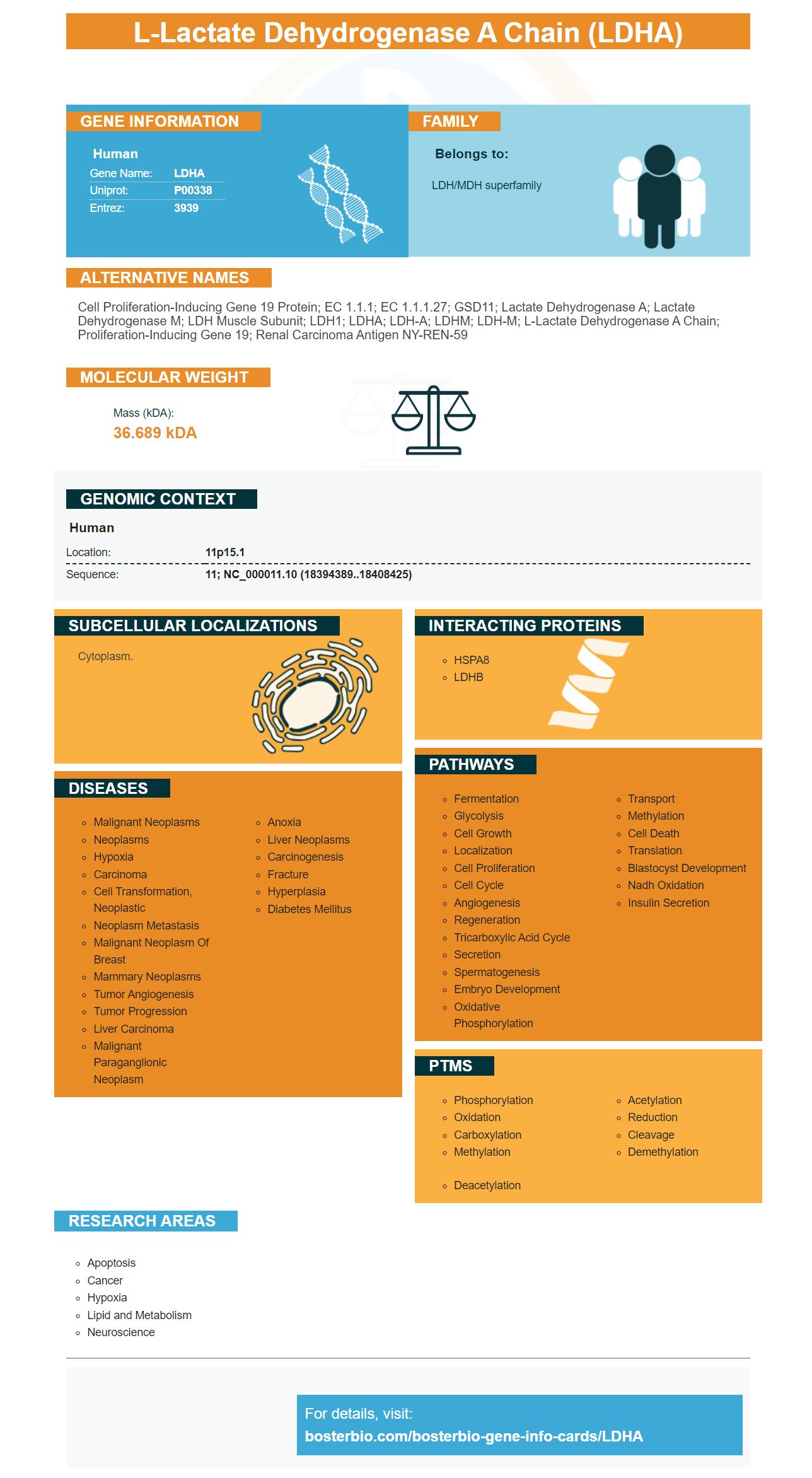
Facts about L-lactate dehydrogenase A chain.

| Human | |
|---|---|
| Gene Name: | LDHA |
| Uniprot: | P00338 |
| Entrez: | 3939 |

| Belongs to: |
|---|
| LDH/MDH superfamily |

Cell proliferation-inducing gene 19 protein; EC 1.1.1; EC 1.1.1.27; GSD11; Lactate Dehydrogenase A; lactate dehydrogenase M; LDH muscle subunit; LDH1; LDHA; LDH-A; LDHM; LDH-M; L-lactate dehydrogenase A chain; proliferation-inducing gene 19; Renal carcinoma antigen NY-REN-59
Mass (kDA):
36.689 kDA

| Human | |
|---|---|
| Location: | 11p15.1 |
| Sequence: | 11; NC_000011.10 (18394389..18408425) |
Cytoplasm.


In this article, we will examine the ways in which LDHA plays a significant role in the progression, metastasis and development of various malignancies, including cancers of the ovarian or breast. This marker also plays an important role in endometriotic and ovarian tissues. Read on to learn more about the most effective uses of this marker. It is a naturally occurring fatty acid that is expressed in various types of tissues.
This protein is a metabolite which aids in the expansion, invasion, and metastasis of many types of cancers. It is increased in tumor cells due to excessively high levels of LDHA. This could be caused by cancer-causing mutations to LDHA's gene. LDHA up-regulation promotes malignant transformation in tumor cells by increasing the production and glycolysis of lactic acids and regulating cancer-related protein. There are promising results from clinical trials focusing on LDHA.
LDHA is mostly found in the cytoplasm, but can also be found in the nucleus or mitochondria. It is a key component of glycolysis and plays an important role likely in DNA duplication or transcription. Studies have demonstrated that LDHA has a high expression profile in various malignancies. This is believed to be due to a variety of mechanisms that influence gene expression.
LDHA is regulated by RCC and has been demonstrated in tumor cell proliferation and survival. LDHA knockdown of cells slows cell growth, migration and inhibits tumorigenesis. In addition, small molecule inhibitors of LDHA reduce the growth of tumor cells in xenograft models. The mechanisms that cause this are still being researched however their importance cannot be overestimated.
In addition to its anti-apoptotic effects, LDHA inhibits EMT by protecting the mitochondria from damage caused by ROS. However, LDHA inhibition also inhibits tumor metastasis. This forces cancer cells to undergo oxidative-phosphorylation to produce ATP. ROS-induced apoptosis is also inhibited by N-acetyl-L-cysteine.
Recent research has proven that LDHA plays a crucial role in the progression, invasion, and development of OSCC. It is crucial to comprehend the role played by LDHA in the progression of this disease because it is involved in the development of tumor cells. Its high level in human OSCC cells has also been proven to influence tumor progression which is the hallmark of the tumor.
Recent research has shown that low levels of LDHA can hinder RCC cell migration in scratch migration assays. These results suggest that LDHA may play a role in the development, invasion, and metastasis in various forms of cancer. These results are in line with the latest research. The treatment LDHA-siRNA inhibits tumor cell migration in the mouse model and could prove to be effective in clinical studies.
Recent research has shown that LDHA expression is very high in the endomometriotic tissue. This may indicate the role played by this protein in the process of developing endometriosis. This article examines LDHA expression in eutopic tissues in order to study the significance of LDHA in endometriotic tissues. However, it's not yet clear what its function is in endometriosis.
To test whether LDHA is involved in the development of endometriosis, researchers knocked down BAX and BAK expression in Ishikawa and THESC cells. These results showed that Ishikawa cells exhibited an increase in the rate of apoptosis after knocking down these proteins. In addition, Western blotting revealed that the knockdown of LDHA caused an increase in expression of proapoptotic proteins.
LDHA regulates glycolysis through catalyzing the conversion of pyruvate to lactic acid. It also regulates the expression and activity of Myc genes, which enhances LDHA. Increased LDHA expression has been linked to metastatic and aggressive cancer. Additionally, it has been linked to endometriosis and non-Hodgkin's lymphoma. This research was made possible by grants from the National Natural Science Foundation of China and the Natural Science Foundation of Zhejiang Province and the Zhejiang Provincial Key Research and Development Program.
Knockdown of LDHA blocks cell migration. It also decreases the survival of cells. It also increases E-cadherin expression and b-catenin expression, while vimentin is the same. These results are in line with previous studies. The LDHA protein is a very abundant protein in endometriotic tissues. It can be helpful in the detection of early signs and symptoms of endometriosis.
Hypoxia is a feature of the endometriotic microenvironment. Hypoxia regulates LDHA expression in tumor cells. In addition, ESCs and immortalized endometrial glandular cells were exposed hypoxia in vitro, and were found to increase the mRNA levels of LDHA and HIF-1a. Hypoxia also blocks cell migration and glycolysis, which may suggest a role for LDHA in the development of endometriotic lesions.
The CDC20 protein controls the cancerous cell cycle and plays a role in metastasis in COAD. Various inhibitors that target CDC20 block the activity of CDC20 which in turn reduces the growth of cancer cells. These include Apcin and Palbociclib. As the findings of this study suggest, LDHA may be an effective therapeutic target in cancer-related chemotherapy.
AMPK signaling causes LDHA in cells. AMPK is involved with the growth and progression of a variety of human cancers. Autophagy deficiency is the most common cause of ovarian cancer. Stress, low cellular energy charge or deprivation can all cause autophagy. LDHA inhibits autophagy and improves the efficacy of the drug vemurafenib (an chemotherapy for ovarian cancer).
Cancer is characterized by reprogramming metabolic processes in tumors. Particularly cancer cells prefer to use aerobic glycolysis to create ATP. This phenomenon is called the Warburg effect. It is caused by lactate dehydrogenaseA. The acidic microenvironment is essential for metastasis. Reduced levels of LDHA reduces the cellular transformation and delays the formation of tumors.
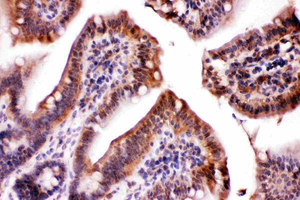
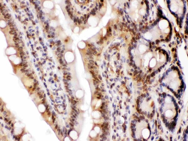
To determine whether LDHA is expressed in ovarian tumors, we performed immunohistochemistry. We employed a conventional immunohistochemical assay. We used an immunohistochemical assay to determine the expression level of LDHA within cancer cells. The cells were stained with an antibody against Annexin V and the data were analyzed using FlowJo software.
In a mouse model of lung metastasis, LDHA knockdown slowed the growth of tumors. Cells from the LDHA knockdown group were found to have smaller tumors and lighter weights than the group that was in control. In addition, LDHA knockdowns led to lower tumor volumes and weights and the tumors that were reduced were found to have fewer proliferating cells. This suggests that LDHA plays a tumor-causing role in PTC cell growth.
A previous study revealed a strong association between LDH levels and the ovarian cancer serum levels. It is possible that the serum LDH can help predict resistance to platinum in patients suffering from cancer of the ovary. LDHA can also be used to develop precision medicine targeted at individual patient's requirements. If this finding is correct, it may be required to include LDHA in blood tests prior to treatment. Next, we will need to find out if LDHA is found in ovarian cancer.
LDHA is an enzyme that plays a crucial role in the glycolysis pathway. It converts pyruvate into lactate, and has been found to be abnormally expressed in breast cancer. It is also associated with malignant tumor growth. Bioinformatic analysis of breast cancer tumors has revealed that LDHA mRNA levels were significantly higher than those of normal breast tissue. The gene POU1F1 also has a significant relationship with LDHA levels of mRNA.
Recent studies have revealed that LDHA is expressed in breast cancers and is associated with tumours that have estrogen receptor. The luminal molecular subtype is associated with LDHA immunoexpression. Higher levels of LDHA could indicate that the cancer cells have a greater proliferative capability. LDHA may also be associated with resistance to hormonal treatment. Furthermore those with breast cancer who have high levels of LDHA also have lower outcomes than BrCa patients.
In vitro kinase tests were carried out by Jin et al. They discovered that Src and Her2 can phosphorylate LDHA at tyrosine 10. These findings are vital to understanding the role LDHA plays in breast cancer metastasis, invasion, and progression. In addition, LDHA can also play an important role in the regulation of metastasis and invasion.
Recent research suggests that LDHA could play a crucial role in RCC metastasis. In the vivo setting, LDHA inhibits tumor metastasis through mechanisms controlled by EMT. In turn, LDHA could be a candidate for RCC treatment. This research has numerous implications for cancer patients and doctors. For example, LDHA may help patients who have invasive bladder cancer.
The role of LDHA in breast cancer remains not known. The breast's ductal area is home to the protein. It is found in tumor tissue at an increased level which suggests that it is present in this tissue. LDHA is an molecule of 37 kD that has anti-tumor effects. It is a biomarker that indicates the aggressiveness of breast cancer. This protein may be useful in developing therapies that target the growth of tumors.
PMID: 3838278 by Tsujibo H., et al. Nucleotide sequences of the cDNA and an intronless pseudogene for human lactate dehydrogenase-A isozyme.
PMID: 3000353 by Chung F.Z., et al. Genomic organization of human lactate dehydrogenase-A gene.
*More publications can be found for each product on its corresponding product page