This website uses cookies to ensure you get the best experience on our website.
- Table of Contents
15 Citations 1 Q&As
16 Citations 1 Q&As
27 Citations
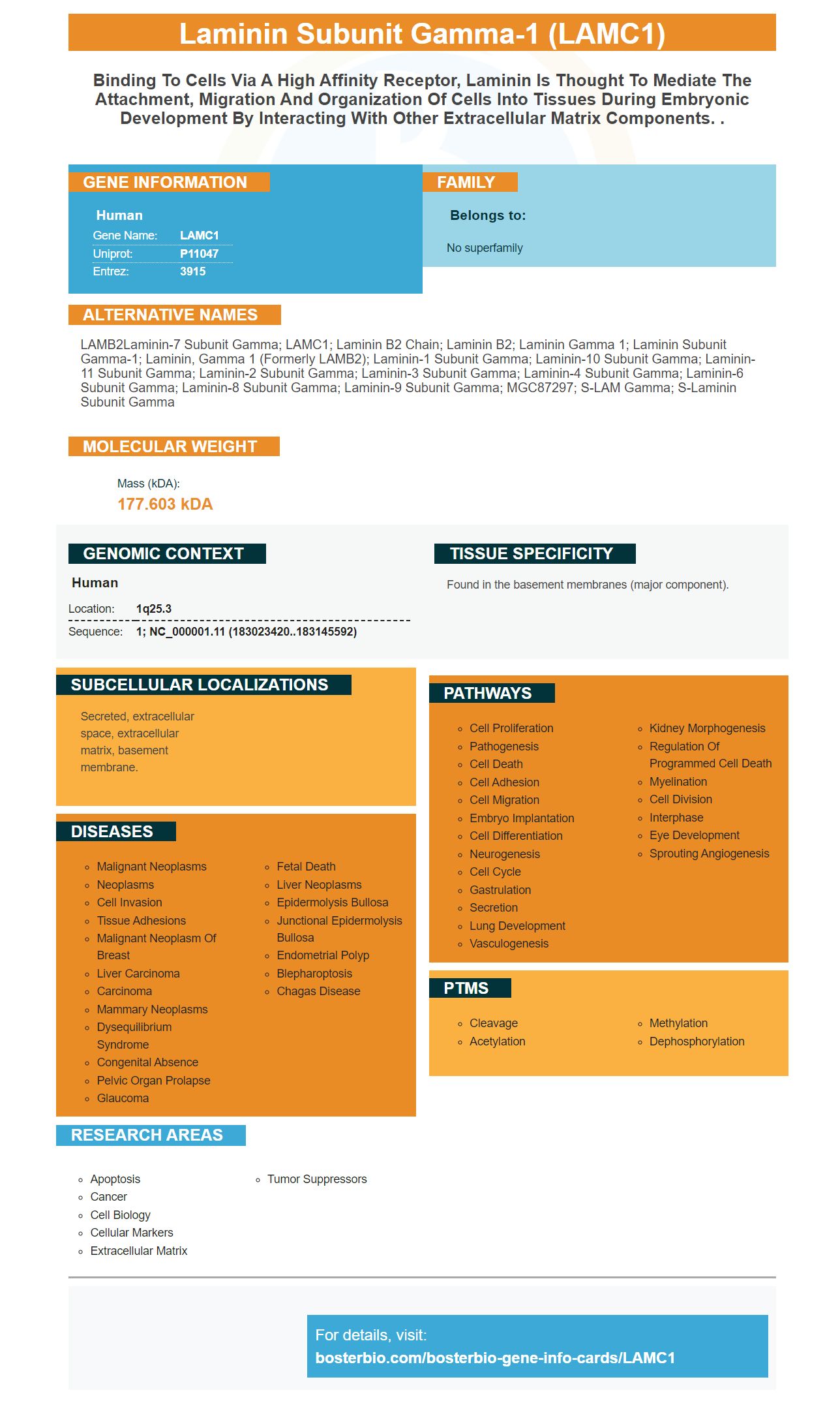
Facts about Laminin subunit gamma-1.
Human | |
---|---|
Gene Name: | LAMC1 |
Uniprot: | P11047 |
Entrez: | 3915 |
Belongs to: |
---|
No superfamily |
LAMB2Laminin-7 subunit gamma; LAMC1; Laminin B2 chain; Laminin B2; Laminin gamma 1; laminin subunit gamma-1; laminin, gamma 1 (formerly LAMB2); Laminin-1 subunit gamma; Laminin-10 subunit gamma; Laminin-11 subunit gamma; Laminin-2 subunit gamma; Laminin-3 subunit gamma; Laminin-4 subunit gamma; Laminin-6 subunit gamma; Laminin-8 subunit gamma; Laminin-9 subunit gamma; MGC87297; S-LAM gamma; S-laminin subunit gamma
Mass (kDA):
177.603 kDA
Human | |
---|---|
Location: | 1q25.3 |
Sequence: | 1; NC_000001.11 (183023420..183145592) |
Found in the basement membranes (major component).
Secreted, extracellular space, extracellular matrix, basement membrane.
The LAMC1Marker is a versatile tool. It can be used for many biological purposes. It can be used for research, as well as for diagnostics and personalized medicine. It is made up of plasma from human beings and is available in many forms. It can be obtained via blood, urine, and feces. Boster has a wide range of products for this purpose, and all of them are backed by extensive research and validation.
To obtain the LAMC1-resistant clone, ES were transformed with clone pCR5, which is G418-resistant. This clone has undergone recombination using the LAMC1 genetic. The internal probe 2 showed a band of approximately 7 kb that was identical to that of the wild-type band. This fragment was then inserted to a KSII Bluescript.
A gene called LAMC1 functions in many cells, and is expressed in many tissues. When expressed in ESCC, it promoted tumor migration and proliferation and was associated with CXCL1 expression. These activities were associated in inflammatory CAF and promoted tumor progression. TGFb upregulation of LAMC1 promotes proliferation, migration, and ESCC cell proliferation, and indirectly induces cancer. LAMC1 could be a prognosis mark for ESCC.
To detect the expression of LAMC1 within cultured cells, PCR was used. The cell medium contains 10% FBS (1% nonessential amino acid) and 1 mm of sodium pyruvate. The BCA Protein Assay Kit was used to determine the protein levels. For detection of LAMC1, RNA was also sequenced.
The duodenal epithelium was and the mesenchyme contained RNA. In the mesenchyme from controls, Lamc1 transcript could be detected. However, it was reduced in mutant mice. Lamc1 knockout mice had no increase in mesenchyme transcripts for laminin-g2 (and alternative laminingamma subunit transcripts), but they were upregulated in epithelial fraction.
40 tumor tissue and 20 normal tissue were taken from the People Hospital LiShui. The samples were frozen in liquid nitrogen to allow for future analysis. All clinical specimens received were consented to by the patient. All research was approved by the Ethics Committee of LiShui University. Following this, RNA samples were taken and detected for the expression of Lamc1 as well as PKM2. The RNA was then used to determine the expression levels of these proteins.
One way to obtain a laminin gene expression level is to infect HepG2 cells with the Lamc1 g2 subunit. These cells are overexpressed for the oePKM2 genes and the results of the infection were quantified at 48 h, 48 h, and 72 hours after Lamc1 infection. Cell proliferation and levels of lactate were also measured. Western blots to measure gene expression were also used.
After preparing the cells in LAMC1mAb, embryoid bodies were fixed with tissue culture plastic. The cells were then allowed to culture for up to 3 weeks. In addition to LAMC1 mAb cells, they also contained myotubes, but displayed no basement membrane. The control cells didn't exhibit any of these properties. These results show that LAMC1 cells can accurately detect this marker when grown in adult tissue cultures.
The best uses of the LAMC1 marker in cancer research are the ability to detect a tumor's mutations before they cause an increase in tumor burden. This gene was found to be associated with low overall survival (OS) and the stage of the tumor in one study. These findings show that the LAMC1 genes is an independent marker for ESCC. Therefore, it may have a useful role in the treatment of ESCC patients.
RT-PCR is one method to determine whether LAMC1 protein exists. The marker is expressed relative the GAPDH control using a lenti ORF-clone. This method can also be used to detect the expression of another gene such as SP. The results are then compared with LO2 in order to determine whether LAMC1 exists.
Although the relationship between LAMC1 gene expression and prognosis of gastric carcinoma is not clear, it appears to have some relevance. It is, in fact, a tumour driver gene. This suggests that it may be prognostic. How do we know if LAMC1 can be used to predict the future? Here are three ways to get LAMC1 for gastric cancer. And as always, keep reading for more information.
The TCGA database includes GC-related gene expression profiles. We used the Rlimma package to identify genes that were differentially expressed. We then used univariate Cox regressions to identify genes associated with survival. ClusterProfiler was also used to analyse pathways and Gene Ontology enrichment for DEGs. Cytoscape was also used to create a protein interaction network that allowed us to identify the LAMC1 gene expression profile. We also validated the LAMC1 expression differential with GEPIA2 & TIMER databases. Finally, we used GSEA and GSVA to determine the major activated pathways.
Lentivirus infection is another way to regulate the expression of LAMC1. SiLamc1-1 and siLamc2 infection in HepG2 cells significantly decreased Lamc1 expression. However, oeLamc1 Lentivirus infection in Huh7 cells increased Lamc1 expression. The use of lentiviruses to test the impact of LAMC1 cells on HCC cells is a useful tool for cancer research.
Our experiments were performed at the Chinese Academy of Medical Sciences, (CAS), and Peking Union Medical College. All experiments were done in accordance with the Animal Care and Use Committees at both institutions. Our experiments were conducted in rooms that had temperatures between 25-27 degrees Celsius and humidity between 45- 50%. In order to establish tumor xenografts in mice, we used a specific technique called ELISA. The Chinese National Cancer Institute used this method extensively.
Anti-LAMC1 antibody has a high specificity for the target subunit. This antibody targets Laminin-521 subunit of laminin. Laminin-521 (a component of basement membrane) is ubiquitously expressed by human pluripotent and stem cells. LAMC1 has three distinct functionally domain structures: the LN and LE domains, and the Cterminal coil-coil fragment. These interactions are vital for the development of several tissues, including the retina and glomerular basement membrane.
PMID: 3360804 by Pikkarainen T., et al. Human laminin B2 chain. Comparison of the complete amino acid sequence with the B1 chain reveals variability in sequence homology between different structural domains.
PMID: 1985895 by Kallunki T., et al. Structure of the human laminin B2 chain gene reveals extensive divergence from the laminin B1 chain gene.
*Showing only the more recent 20. More publications can be found for each product on its corresponding product page