This website uses cookies to ensure you get the best experience on our website.
- Table of Contents
1 Citations 3 Q&As
Facts about Lysophosphatidic acid receptor 6.
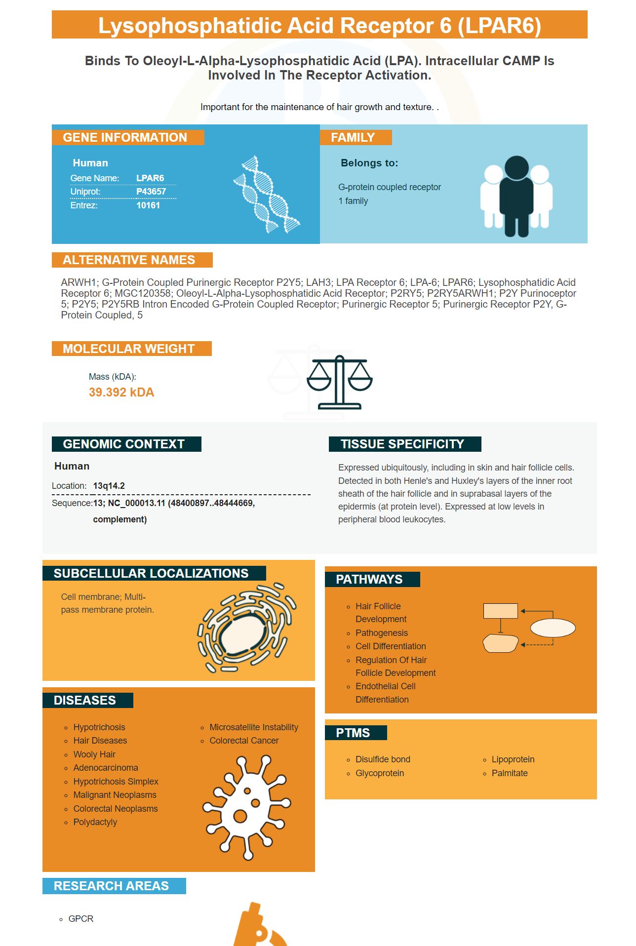
Important for the maintenance of hair growth and texture. .
Human | |
---|---|
Gene Name: | LPAR6 |
Uniprot: | P43657 |
Entrez: | 10161 |
Belongs to: |
---|
G-protein coupled receptor 1 family |
ARWH1; G-protein coupled purinergic receptor P2Y5; LAH3; LPA receptor 6; LPA-6; LPAR6; lysophosphatidic acid receptor 6; MGC120358; Oleoyl-L-alpha-lysophosphatidic acid receptor; P2RY5; P2RY5ARWH1; P2Y purinoceptor 5; P2Y5; P2Y5RB intron encoded G-protein coupled receptor; Purinergic receptor 5; purinergic receptor P2Y, G-protein coupled, 5
Mass (kDA):
39.392 kDA
Human | |
---|---|
Location: | 13q14.2 |
Sequence: | 13; NC_000013.11 (48400897..48444669, complement) |
Expressed ubiquitously, including in skin and hair follicle cells. Detected in both Henle's and Huxley's layers of the inner root sheath of the hair follicle and in suprabasal layers of the epidermis (at protein level). Expressed at low levels in peripheral blood leukocytes.
Cell membrane; Multi-pass membrane protein.
If you want to submit results of your special samples, species, applications, and other research to receive product credits, you may want to do so using Boster Bio. This application is open to all scientists worldwide. This article outlines how scientists can use this LPAR6 marker for their research. It also covers how LPAR6 is downregulated in TCGA and LUAD cells.
High mRNA expression of LPAR6 was associated with a better prognosis in patients with ACC, LGG, and SKCM. However, expression was decreased in COAD, BRCA, HNSC, LUAD, and READ. These data suggest that LPAR6 expression might be associated with poorer prognosis in other cancer types.
Although few studies have shown the function of LPAR6, many have indicated that it plays a significant role in the immune system. Moreover, its association with tumor infiltration and prognosis is unclear. The present study analyzed mRNA expression of LPAR6 across multiple databases and evaluated its association with immune infiltrates. In addition, immunohistochemistry was performed for LPAR6 in lung squamous cell carcinoma and adenocarcinoma. Based on these findings, a systematic prognostic landscape was constructed.
The study also examined the relationship between LPAR6 mRNA expression and patient survival in various types of cancers. The study also examined whether higher LPAR6 mRNA expression was associated with improved survival in lung cancer patients. Although a number of factors could influence LPAR6 expression, the findings have some clinical relevance to lung cancer. In the meantime, more research is needed to better understand the role of LPAR6 in different cancer types.
Interestingly, LPAR6 is found to negatively correlate with tumor purity. The level of mRNA encoding LPAR6 was positively correlated with immune infiltrating cells in LUAD. These findings suggest that LPAR6 is associated with improved survival in patients with LUAD. It may also play an important role in embryonic angiogenesis. The results of this study are promising and suggest that LPAR6 can help patients with LUAD find the optimal treatment.
LPAR6 is expressed in more advanced stages of gliomas than in the general population. The ATX-LPAR pathway contributes to the progression of the cancer by regulating the expression of the ATX/LPA pathway. Several studies have suggested that the LPAR1-PKCa pathway may help in the reversal of pathologic responses. This study has also found that hyperglycemia increases the expression of VEGF-C. The multifunctional chaperon protein calreticulin may modulate this process.
LPAR6 has a broad cellular function. It has biological functions in the central nervous system. In addition to activating various types of T cells, it also inhibits the growth of cancer cells. In addition to its cytotoxicity, LPAR6 is involved in the control of inflammation and autoimmune diseases. In addition, LPAR6 inhibits tumor cell migration in colorectal cancer.
LPAR6 expression levels were higher in certain types of cancer than in others. Cancers of the brain, central nervous system, liver, kidney, pancreatic, and prostate were more likely to express LPAR6 than other types of tumors. However, the level of LPAR6 was lower in other types of tumors and adjacent normal tissues. In addition, LUAD and LUSC expressed LPAR6 in different amounts.
LPAR6 is found to be expressed on the surface of different types of T cells, including CD4+ and CD8+. LPAR6 has been associated with the infiltrating level of CD8+ T cells, CD4+ T cells, and dendritic cells. In addition, the presence of LPAR6 in tumors can regulate the depolarization of macrophages in the tumor microenvironment. Furthermore, it may serve as a novel target for LUAD therapy.
Researchers have been using RNA-Seq to identify how LPAR6 affects the prognosis of different types of cancer. For instance, higher expression of LPAR6 was associated with a better prognosis in liver cancer. Further, higher LPAR6 protein levels were associated with better overall survival in LUAD and LUSC cancer patient cohorts. So, the gene may have a specific role in predicting survival in lung cancer.
The expression of LPAR6 in tumors correlates with the immune-infiltrating cells in LUAD. LUAD patients with higher levels of LPAR6-positive T cells have better prognoses. This may be due to interactions between LPAR6 and immunocytes. The study suggests that LPAR6 is essential for proper immune cell recruitment in LUAD.
Furthermore, the LPAR6 gene is also required for proper T-cell motility in confined 3D environments. The LPA2-LPA2 axis is a key determinant in the control of T-cell motility in LN parenchyma. The gene's function is not fully understood, but it plays a crucial role in regulating T-cell motility and adhesion.
Researchers have found that LPAR6 is downregulated in the tumor tissues of LUAD patients and that its mRNA expression correlates with patient OS. Moreover, LPAR6 has been specifically correlated with immune infiltrating cells in LUAD, suggesting that LPAR6 may play a role in the recruitment of these immune cells. Nevertheless, further studies are required to determine whether this protein is indeed responsible for regulating the immune response in LUAD.
The association between LPAR6 expression and tumor immune responses was examined in a large cohort of LUAD patients. In addition, the expression level of LPAR6 was correlated with immune cell counts in LUAD tumors, whereas it was unrelated in LUSC patients. However, these findings suggest that LPAR6 may be a promising novel biomarker of LUAD treatment.
Interestingly, LPAR6 is associated with a good prognosis in some types of cancer, as it is associated with the recruitment of peritumoral fibroblasts. Moreover, LPAR6 has been found to be involved in immunotherapy, a new genre of cancer treatments. The tumor microenvironment plays an important role in this form of therapy. So, it is imperative to understand and treat this cancer with immunotherapy.
The analysis of LPAR6's association with lung cancer progression involved several different bioinformatics methods. A network of 19 proteins co-expressed with LPAR6 was constructed to determine LPAR6's potential interactions with cancer-associated proteins. The interactions between LPAR6 and these 19 proteins were examined using Pearson's correlation coefficient (R-squared) and the network's top 50 positively and negatively correlated genes.
Furthermore, lower levels of LPAR6 were negatively associated with both lymph node metastasis and clinical stage of the cancer. This further highlights how LPAR6 may play a role in the treatment of breast cancer. It is possible to use this information to target LUAD with targeted therapy. But what exactly is LUAD? And what exactly does it do? Well, it affects tumors in a wide variety of ways.
It is important to note that CLEC9A is expressed in a variety of human LUAD cell lines. Human LUAD cell lines with the highest and lowest levels of CLEC9A expression were studied. Moreover, pcDNA3.1-CLEC9A constructs and CLEC9A siRNA were used to control endogenous expression of CLEC9A in the NCI-h2395 cell line. Transfection of the pcDNA3.1-CLEC9A construct with NCI-h2395 cells decreased the level of CLEC9A in LUAD cell lines.
TCGA data show that LPAR6 is downregulated in a subset of breast cancers. The LPAR6 gene, which encodes a protein that controls cell proliferation, is related to the E2F and RB1 families. In several studies, LPAR6 expression level has been associated with a favorable prognosis. However, the mechanisms behind this association remain unclear.
In a recent study, LPAR6 expression was examined in a group of cancers, using RNA-Seq data in TCGA. Overall, LPAR6 was expressed at lower levels than in normal tissues. In addition, LPAR6 expression levels were different among different types of cancer. In a study, LPAR6 expression was found to be inversely related to clinical stage and lymph node metastasis, suggesting a role for LPAR6 in cancer development.
Moreover, LPAR2 is associated with immune cell infiltration in HNSC and KIRC. It was significantly correlated with infiltrating B cells, CD4 + T cells, neutrophils, and DCs. These data indicate that LPAR2 plays a pivotal role in the immune microenvironment of tumors. But further studies are needed to elucidate the role of LPAR2 in the immune system.
Several functional cells are associated with LPAR6 expression, including monocytes and T cells. In addition, the expression of LPAR6 has been found to be associated with M1 and M2 macrophages, which are responsible for preventing tumor development. Additionally, the expression of LPAR6 in tumor tissue shows a strong correlation with NOS2 and PTGS2, which are two major immune cell markers. These findings suggest a possible role for LPAR6 in tumor growth.
Expression levels of LPAR6 were positively correlated with levels of CD4+ T cells in 24 types of cancers. In addition, LPAR6 was positively associated with levels of neutrophils, dendritic cells, and macrophages in 23 types of cancer. However, these data are not predictive of survival. This is in part because patients with tumors with higher LPAR6 levels had poorer outcomes.
The TCGA data are from over 10,000 patients. It contains information on 32 types of cancer and a meta-analysis of LPAR6 expression in those groups was carried out using LinkedOmics. Using a statistical tool called LinkFinder, we identified genes with differential expression levels in TCGA. We then used Pearson's correlation coefficient to investigate correlations between gene expression levels and survival. We also used LinkInterpreter to identify networks and pathways, which was highly predictive of survival.
PMID: 7902321 by Toguchida J., et al. Complete genomic sequence of the human retinoblastoma susceptibility gene.
PMID: 8889552 by Herzog H., et al. Intron 17 of the human retinoblastoma susceptibility gene encodes an actively transcribed G protein-coupled receptor gene.
*More publications can be found for each product on its corresponding product page