This website uses cookies to ensure you get the best experience on our website.
- Table of Contents
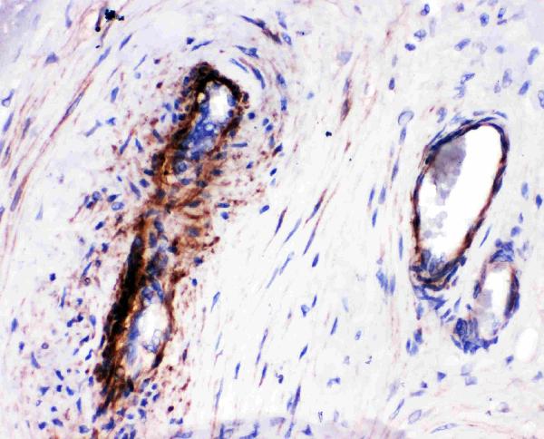
1 Citations 4 Q&As

Facts about LIM and SH3 domain protein 1.

. .
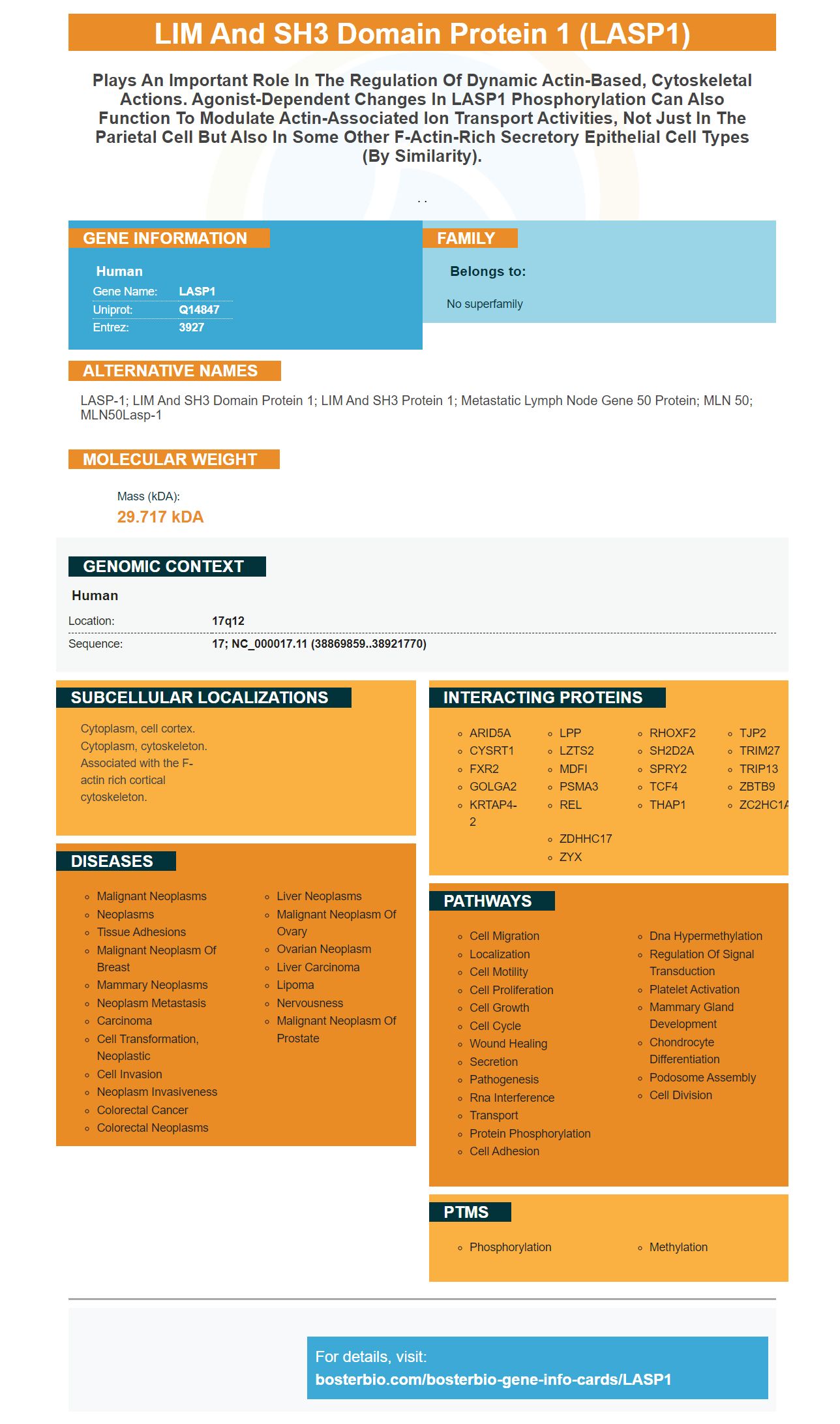
| Human | |
|---|---|
| Gene Name: | LASP1 |
| Uniprot: | Q14847 |
| Entrez: | 3927 |

| Belongs to: |
|---|
| No superfamily |

LASP-1; LIM and SH3 domain protein 1; LIM and SH3 protein 1; Metastatic lymph node gene 50 protein; MLN 50; MLN50Lasp-1
Mass (kDA):
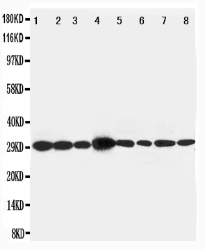
29.717 kDA

| Human | |
|---|---|
| Location: | 17q12 |
| Sequence: | 17; NC_000017.11 (38869859..38921770) |
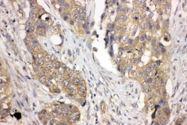
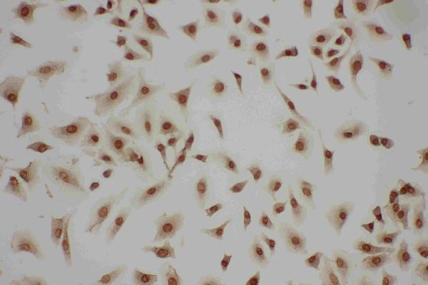
Cytoplasm, cell cortex. Cytoplasm, cytoskeleton. Associated with the F-actin rich cortical cytoskeleton.


PMID: 7490069 by Tomasetto C.L., et al. Identification of four novel human genes amplified and overexpressed in breast carcinoma and localized to the q11-q21.3 region of chromosome 17.
PMID: 7589475 by Tomasetto C., et al. Lasp-1 (MLN 50) defines a new LIM protein subfamily characterized by the association of LIM and SH3 domains.
*More publications can be found for each product on its corresponding product page

