This website uses cookies to ensure you get the best experience on our website.
- Table of Contents
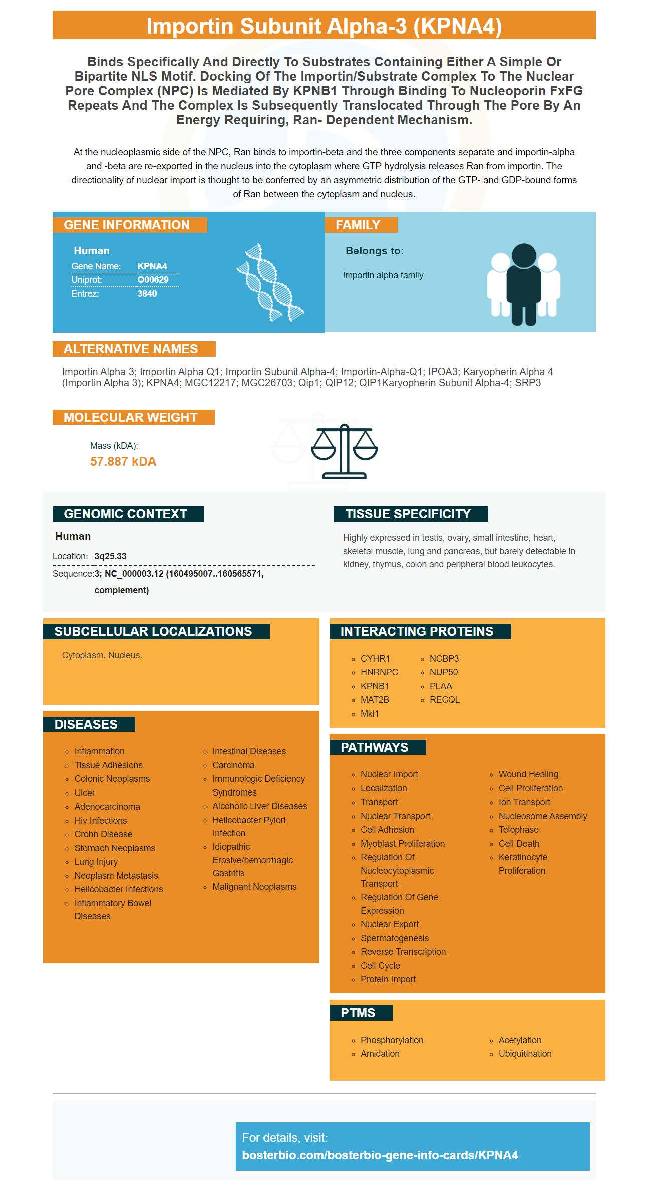
Facts about Importin subunit alpha-3.
At the nucleoplasmic side of the NPC, Ran binds to importin-beta and the three components separate and importin-alpha and -beta are re-exported in the nucleus into the cytoplasm where GTP hydrolysis releases Ran from importin. The directionality of nuclear import is thought to be conferred by an asymmetric distribution of the GTP- and GDP-bound forms of Ran between the cytoplasm and nucleus.
Human | |
---|---|
Gene Name: | KPNA4 |
Uniprot: | O00629 |
Entrez: | 3840 |
Belongs to: |
---|
importin alpha family |
Importin alpha 3; Importin alpha Q1; importin subunit alpha-4; importin-alpha-Q1; IPOA3; karyopherin alpha 4 (importin alpha 3); KPNA4; MGC12217; MGC26703; Qip1; QIP12; QIP1Karyopherin subunit alpha-4; SRP3
Mass (kDA):
57.887 kDA
Human | |
---|---|
Location: | 3q25.33 |
Sequence: | 3; NC_000003.12 (160495007..160565571, complement) |
Highly expressed in testis, ovary, small intestine, heart, skeletal muscle, lung and pancreas, but barely detectable in kidney, thymus, colon and peripheral blood leukocytes.
Cytoplasm. Nucleus.
Boster is the ideal place to find primary antibodies that detect this specific protein. Their antibodies are high-affinity and have been extensively validated in Western blotting, Immunohistochemistry, and ELISA. This gives you confidence in their high-quality primary antibodies as well as their performance in a wide array of applications. Below are details about Boster’s antibodies for the KPNA4 indicator.
Boster has high affinity primary antibodies which are highly correlated to human KPNA4 gene expression. These antibodies have been validated by Western Blotting, Immunohistochemistry, and ELISA. They are therefore an excellent choice for research projects. They are compatible for the KPNA4 gene marker and have a high citation rate in the scientific literature. They are a reliable source to primary antibodies that are widely used for cell biology.
Boster Bio KPNA4 marker Western Blot Antibody detection method can be used to determine protein expression in cell samples. Both qualitative and quantitative Western blot techniques are available. A negative control (lysate from a cell without protein expression) is used to ensure antibody specificity. The secondary antibody is used for detection to determine the binding and blocking capabilities of the primary antibody. To ensure that the secondary antibodies system is working properly, it is necessary to use a blank control and a load control in addition to detecting the protein.
To determine the level of protein expression, a membrane stained using Ponceau S containing 0.2% W/V glacial Acetic Acid can be used. This will allow you to determine the transfer efficiency. A constant current of 150-300 mA is usually applicable for this procedure. The gel's concentration is also important in determining the protein transfer efficiency. The longer the transfer process takes, the higher the gel concentration.
Western blotting also known by protein immunoblotting is a process that detects certain proteins in a sample. The process consists of five steps: SDS-polyacrylamide gel electrophoresis, transfer, blocking, and secondary antibody incubation. The detection method is extremely sensitive and specific. Western blotting is a widely used method in molecular biology, biochemistry and immunogenetics.
Boster Bio KPNA4 marker Western Blot detection tools include two types controls. Electrophoresis can be used to confirm the Northern Western Blot. The Southern Western Blot antibody detection method involves membrane discovery. This multi-step process requires expertise and experience. There are many steps involved with Western blotting. Each step can introduce considerable variability.
The ECL chemiluminescent detector kit is designed for detection of protein expression on membranes. It is also capable of multiplex-blotting. The ECL Western Blotting System is optimized for single and multiplex detection of protein using nitrocellulose. Hybond ECL membranes are 0.45 pore in size, which gives them excellent sensitivity and low background. They are compatible to most buffers and nitrocellulose membranes.
Chemiluminescent western Blotting starts with a primary antibody that recognizes target protein. The secondary antibody is then labeled with an enzyme, such as horseradish Peroxidase, alkaline Phosphatase or luminol-based HRP. The chemiluminescent signal that results is measured using either an x-ray film, or digital imaging.
The Clarity Max ECL ECL-chemiluminescent detection systems can be used with any horseradish peroxidase conjugate. The Clarity Max is ideal for western blot applications. The Clarity reagents can be used with any horseradish peroxidase conjugate, including anti-ceruloplasmin and anti-transferrin antibodies.
The ECL chemiluminescent detect kit's most important feature is its ability to detect the expression of protein. The technology uses bovine IgG–HRP, 0.1% Tween-20 and PBS-Tris pH 7.4 (22 degC). The ECL Chemiluminescent Detection System will not function if the reagents cannot be mixed in equal amounts.
A DAB-chromogenic detection system can be used to detect DNA. This system utilizes a proprietary DNA chromogenic reagent XLGLRWLLLVXVLKGVQCQSLEXSGGRDOWDETL. The enzyme DAB is capable of detecting DNA strands that have over 90% identity. The system uses a chromogenic counterstain, Methyl Green. This solution contains 0.125% VWR powder and 0.1M sodiumcetate buffer (pH 4.2-4.3). The tissue is mounted to a slide the day before. The slides are then dehydrated in acetone or with distilled waters and processed through a series on ethanol steps to remove the counterstain. The Methyl Green counterstain in the cortex is more nuclear.
This kit allows the extraction of nuclear protein from fresh tissues or cultured cells. KPNA4 proteins are stable at 4degC. They can also be shipped at ambient temperature. The kit contains all the reagents you need to separate nuclear protein from fresh tissues and cultured cells. This kit is able to distinguish between cytoplasmic protein and nuclear proteins, in addition to offering a high sensitivity ELISA test kit.
PMID: 9168958 by Seki T., et al. Cloning of a cDNA encoding a novel importin-alpha homologue, Qip1: discrimination of Qip1 and Rch1 from hSrp1 by their ability to interact with DNA helicase Q1/RecQL.
PMID: 9395085 by Koehler M., et al. Cloning of two novel human importin-alpha subunits and analysis of the expression pattern of the importin-alpha protein family.