This website uses cookies to ensure you get the best experience on our website.
- Table of Contents
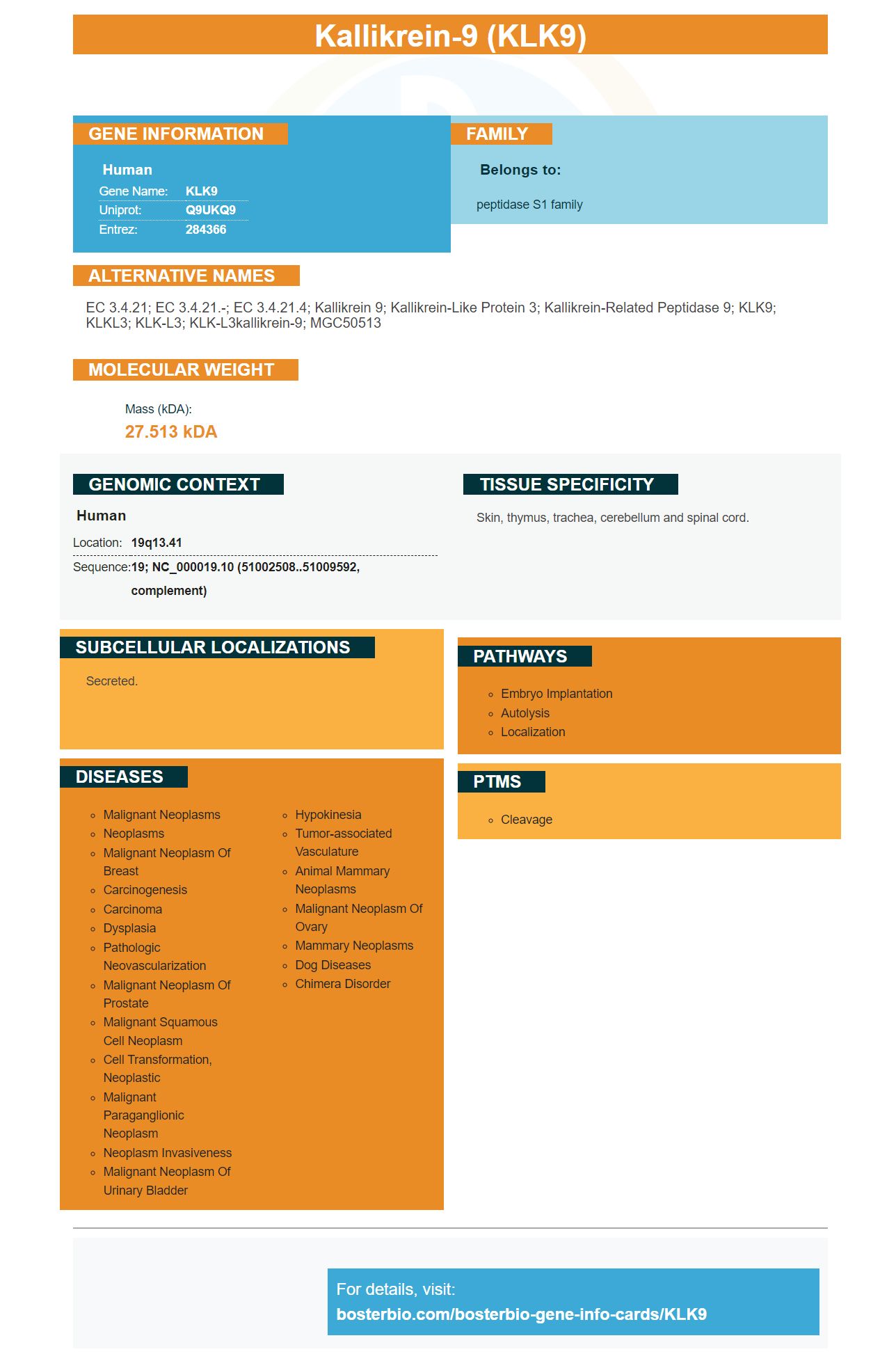
Facts about Kallikrein-9.
Human | |
---|---|
Gene Name: | KLK9 |
Uniprot: | Q9UKQ9 |
Entrez: | 284366 |
Belongs to: |
---|
peptidase S1 family |
EC 3.4.21; EC 3.4.21.-; EC 3.4.21.4; Kallikrein 9; Kallikrein-like protein 3; kallikrein-related peptidase 9; KLK9; KLKL3; KLK-L3; KLK-L3kallikrein-9; MGC50513
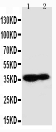
Mass (kDA):
27.513 kDA
Human | |
---|---|
Location: | 19q13.41 |
Sequence: | 19; NC_000019.10 (51002508..51009592, complement) |
Skin, thymus, trachea, cerebellum and spinal cord.
Secreted.
A biotinylated KLK9 mark has many advantages. The antibodies are very specific and can be used for assays to detect heterocomplexes with KLK9 and serine protease inhibitors. Boster is also a world leader in biotinylation. Because you can create your own antibodies, without needing any expertise or specialized equipment, this technology is a game changer.
To determine the concentrations of Biotinylated mak-KLT9 within the assay diluent samples and standards were diluted in 6% BSA. Next, 50 mL of assay buffer A was added to each well. Buffer A contains 60 g/L BSA, 25 mL/L normal mouse serum or goat serum, 10 g/L bovine IgG, and 50 mM Tris-HCl pH 7.8. The buffer also contains 0.005% of Tween-20.
Each well was then infused with 100 microliters (pH 7.8) of 50mM Tris buffer. The wells were then incubated for 2 h at RT. After that, alkaline phosphatase-conjugated streptavidin was added to each well. The plate was then rinsed three times with PBS and 50 mg ABC.
A solution of biotinylated Mat-KLK9. The spacer arm between biotin (the reactive moiety) and the four-ethylene glycol chains was then conjugated in this assay. The assay diluent then disperses biotinylated mat KLK9. A biotinylated matrix-KLK9 reagent is an excellent choice for protein-based analysis.
It is simple to conduct a cell-surface Biotinylation Assay. It involves labeling target proteins with biotin. The biotinylated mat-KLK9 is attached to beads coated with streptavidin, which has a high affinity for biotin. The beads that have been biotin-coated are removed in a subsequent step. Western blotting is a method to visualize the target proteins. The percentage of endocytosed is determined by the density and color of the bands.
The SRM technique for quantitative analysis KLK9 in BosterBio is a two step process. Firstly, mat-KLK9 was purified using anion-exchange chromatography (AEC) and diluted three times with equilibration buffer. Second, Mat-KLK9 has been purified using reversed-phase HPLC. Third, the fractions containing KLK9 were mixed with 1% TFA and loaded onto a Restek column (Viva C4 series).
This experiment used the SRM method to quantitative KLK9 to analyze matKLK9 protein in suspension cells. The cells were able to produce 10-11 mg/L of protein 96 hours after transfection. The protein was analyzed by using a heavy-labeled peptide, which migrated at approximately 42 kDa. PNGaseF was used to reduce the 42-kDa band from about 25 KDa. The protein was then isolated on a large scale.
The SRM method for quantification of KLK9. The SRM technique was applied to the quantification of KLK9. The newly developed 28ED436 anti-mouse antibody was used to validate and validate the SRM technique. All monoclonal antibodies were tested for repeatability. The light/heavy ratio of monoclonal antibodies was also evaluated. It was approximately 13% in the case of antibodies with higher affinity and 20% in spiked heavy peptides.
The SRM method to measure KLK9. ELISAs can be used to determine whether KLK9 is bound or free. Researchers have used the SRM method to determine KLK9 in BosterBio ELISA for over a decade. It will likely be used in clinical trials to identify disease biomarkers.
We performed a reverse -phase HPLC ELISA in order to determine the KLK9 content of a sample. The assay was performed on a white polystyrene microtiter plate coated with 500 ng of mouse monoclonal anti-KLK9 antibody. This was then diluted with 50mM Tris buffer, 150mM NaCl. The eluates of the mixture were then analysed using western blotting and SRM. The purity of the samples was confirmed by Coomassie and silver staining.
Three mAbs, pro-KLK9 and mature KLK9, were tested during the ELISA. The assay used mature KLK9, whereas pro KLK9 could be purchased from R&D Systems. Two of the mAbs used were 28ED436, and 4ED28.
For the assay, antibodies against KLK9 was prepared by adding Buffer B to wells. The second step involved the addition of polyclonal antibodies to serine protease inhibitors. These were antithrombinIIIpAb and alpha-2–antiplasmin antibodies, as well as anti-chymotrypsin. A control was added to the biotinylated anti-KLK9 antibody.
After the assay was completed, the peptides were examined for their cytotoxicity against J774.2 mouse mammalian macrophages. The peptides proved to be effective vaccine candidates against Schistosomiasis after three days. 90% of the macrophages survived after three days. These assays enable scientists to monitor KLK9 levels at various clinical settings.
LifeTechnologies developed the Expi293 mammalian expression system to increase the number of gene-expressed protein in mammalian cells. Compared to HEK293 cells, Expi293 cells grow denser in culture and produce a higher expression yield. LifeTechnologies recommends transfection using ExpiFectamine. However, this reagent can be expensive. Alternatively, PEI, which is 1/1000 the cost, can be transfected with Expi293 cells. PEI can also be used to generate multiple versions of tetravalent, bispecific antibodies.
The Expi293 mammalian expression system is a good choice for transient high-yield protein generation. Its unique transfection process uses high-density cell cultures 293F to produce expression levels of human proteins exceeding 1 g/L. This new technology enables the production of proteins in very small cultures and yields up to 2-10 fold more proteins than previous generations of transient expression systems.
This system has a major advantage: it requires less DNA and other reagents than other expression systems. This makes it a cost-effective solution for high-density mammalian cell cultures. The Expi293 mammalian expression system for protein expression is compatible with many applications, and highly customizable. It does not perform as well HEK293 cells.
Expi293 cells have a faster growth rate than other HEK293 lines. They also require less medium and handling. Expi293F cells are faster than the predecessor cell line and can reach high cell densities. They can also express proteins more efficiently than HEK293 and Expi293F cells. This cell line can be used for PEI transfection, which is a simple and reliable way to produce large amounts protein.
The Boster Bio mouse anti-KLK9 monoclonal antibody (mAb) is used for detection and capture of KLK9. It was diluted in 50 mM Tris buffer, pH 7.8, and then added to a 96-well polystyrene microtiter plate. Each well was coated with 100uL matKLK9-antigen diluted in PBS (1% BSA). Negative controls were used for wells that did NOT contain antigen. After coating the plate with PBS, the plates were rinsed three times with 50 mM ABC.
The boster-bio mouse anti KLK9 antibody has been validated in multiple studies using a variety different methods. This antibody was also validated in Western Blotting. The Boster anti-KLK9 mAb can also recognize a number of other proteins in the body. The company also manufactures a rabbit anti-KLK9 mAb that is specific to KLK9, as well as a mAb that targets other proteins.
To create Boster Bio mouse anti-KLK9 antibodies, a mature form of KLK9 was expressed in Expi293 mammalian cell culture. Gene Art created a plasmid containing both the KLK9 and IgK chains secretion signal proteins. Next, E. coli TOP10 chemically competent cells amplified the pCDNA3.4-KLK9 plasmid. The PureLink (tm), HiPure HiPure DNA midiprep kit purified the plasmid.
Boster Bio pCDNA3.4 KK9 plasmid will provide you with a high-quality plasmid. It has binding activity to KLK9! The binding protein can be bound to a variety targets antigens such as interleukin 13 or prostaglandin E2, interleukin 18, and interleukin 18. It can also bind the cellular proteins Factor VII and Factor IX. Many Boster Bio's pCDNA3.4KLK9 kits have been tested against more than 250 tissues. These antibodies are extremely specific, which results is high affinity and precision.
PMID: 10652563 by Yousef G.M., et al. Identification of novel human kallikrein-like genes on chromosome 19q13.3-q13.4.
PMID: 10783266 by Yousef G.M., et al. The expanded human kallikrein gene family: locus characterization and molecular cloning of a new member, KLK-L3.