This website uses cookies to ensure you get the best experience on our website.
- Table of Contents
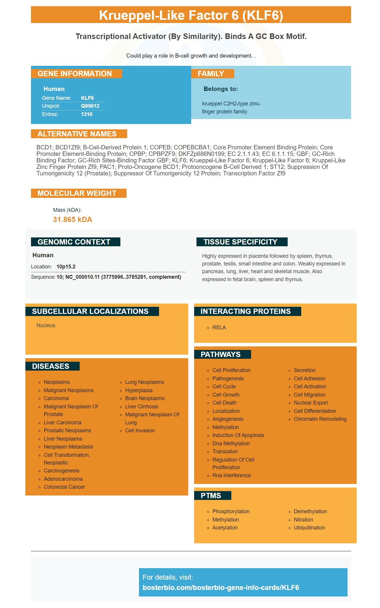
Facts about Krueppel-like factor 6.

Could play a role in B-cell growth and development. .
| Human | |
|---|---|
| Gene Name: | KLF6 |
| Uniprot: | Q99612 |
| Entrez: | 1316 |

| Belongs to: |
|---|
| krueppel C2H2-type zinc-finger protein family |

BCD1; BCD1Zf9; B-cell-derived protein 1; COPEB; COPEBCBA1; core promoter element binding protein; Core promoter element-binding protein; CPBP; CPBPZF9; DKFZp686N0199; EC 2.1.1.43; EC 6.1.1.15; GBF; GC-rich binding factor; GC-rich sites-binding factor GBF; KLF6; Krueppel-like factor 6; Kruppel-like factor 6; Kruppel-like zinc finger protein Zf9; PAC1; Proto-oncogene BCD1; protooncogene B-cell derived 1; ST12; suppression of tumorigenicity 12 (prostate); Suppressor of tumorigenicity 12 protein; Transcription factor Zf9
Mass (kDA):
31.865 kDA

| Human | |
|---|---|
| Location: | 10p15.2 |
| Sequence: | 10; NC_000010.11 (3775996..3785281, complement) |
Highly expressed in placenta followed by spleen, thymus, prostate, testis, small intestine and colon. Weakly expressed in pancreas, lung, liver, heart and skeletal muscle. Also expressed in fetal brain, spleen and thymus.
Nucleus.


The function of KLF6 is largely unknown. This article will examine the role of KLF6 in odontogenesis and iMDP-3. It will also provide insight into the role of KLF6 in regulating p21 expression. Boster Bio: Best Uses Of The KLF6 Marker
The p21 protein is an important regulatory factor that controls cell cycle progression. It is also involved in suppressing the functions of the tumor suppressor gene p53. This research indicates that KLF6 regulates p21 expression. However, it remains unclear whether KLF6 affects the expression of p21 in cancer cells. For now, however, this research provides a new insight into how KLF6 regulates p21 in Boster Bio.
The peptides containing KLF6 were synthesized by combining four peptides available commercially. These peptides covered lysine residues in the transactivation domain and DNA-binding domain, respectively. The peptides were then analyzed by MALDI-MS with a-cyano-4-hydroxy-cinnamic acid as the matrix. The fragment spectra revealed the acetylation sites at each lysine residue.
It is currently unclear if KLF6 directly inhibits apoptosis, but it does seem to influence the expression of p21 in cells. The increased expression of KLF6 in Boster Bio cells is associated with apoptosis resistance. Therefore, there may be some correlation between KLF6 expression and resistance to chemotherapy. Moreover, KLF6 is associated with cancer-related signaling pathways, such as apoptosis and cell migration.
The activities of KLF6 alter the levels of p21WAF1 and cyclin D1 in cells. KLF6 activity promotes G1 cell cycle arrest and alters p21WAF1/cip1 expression. In Boster Bio cells, KLF6 also promotes cell differentiation and sequestration through acetylation of its lysine residue K209.
The expression of p21 was assessed in fifteen healthy people and fifteen esophageal cancer patients. Interestingly, the levels of p21 were not significantly different in the two groups of patients. Further, the expression of p21 was also not significantly different between males and females. Furthermore, the results did not show a correlation between p21 expression and tumor prognosis.
Transient transfection of a plasmid encoding KLF6 was performed using Lipofectamine 2000 reagent from Invitrogen. The transfected PC3M cells were cultured in 10 cm dishes and lysed 36 hours later. The cells were co-transfected with a puromycin-expressing plasmid in a 10:1 ratio. Puromycin-expressing luciferase assay was conducted on these transfected cells.
To determine if KLF6 is involved in p21 expression in Boster Bio, amplification of the p21 gene was conducted in cells. The temperature used to maximize the transcription of the p21 gene was 58degC. The Ct values for each sample were calculated, and the amount of fluorescent light used was subsequently measured. The corresponding p21 gene expression was then measured by GAPDH.
In murine odontoblasts, the expression of KLF6 and P21 were found to be co-expressed during tooth development. Both proteins are expressed in the nucleus and cytoplasm of iMDP-3 cells. Co-expression was also confirmed using RT-qPCR. These results suggest that KLF6 and P21 are involved in odontoblast differentiation.
The expression levels of KLF6 in the tooth germs can contribute to our understanding of odontogenesis. The expression level of KLF6 in the tooth germ may also help clarify its association with odontoblastic differentiation. We have previously found that KLF6 was expressed in several cell types during tooth germ development, and H&E staining revealed that P21 was also expressed in various types of tooth germs.
The expression and function of these factors during tooth development has been demonstrated in several studies. The aim of this review is to synthesize all known information on KLF6 in odontogenesis. During tooth development, the first morphological signal of tooth formation is the appearance of epithelial thickenings into ectomesenchyme at sites that correspond to presumptive teeth. The number of thickenings in mice is lower than that in humans, who have three molars separated by diastema regions.
The expression of C5L2 and KLF6 in odontoblasts is required for tooth development. This gene is also important in the Wnt1/b-catenin signaling pathway and is implicated in odontoblast differentiation. The function of this gene in tooth development remains unclear. In addition to regulating tooth growth, the gene also controls the differentiation of hDPSCs into odontoblasts.
MiR-148a-3p plays an important role in cell proliferation, invasion, and differentiation. It modulates Wnt1 signaling and hDPSC differentiation to odontoblasts. However, it has not been fully understood how these genes interrelate with one another. Interestingly, KLF6 and miR-148a-3p play similar roles in bone and tooth development.
To study KLF6's regulatory effects on iMDP-3 cells, we transfected iMDP-3 cell lines with flag-hKLF6 or control plasmids. Twenty-four hours later, we harvested the cells and performed RT-qPCR and western blotting to determine protein expression. Afterward, we performed flow cytometry analysis and ChIP to identify whether KLF6 overexpression had any effect on apoptosis in iMDP-3-expressing cells.
Previous studies have shown that claudin family members exhibit spatiotemporal gene expression patterns during tooth development. In particular, Cldn10 promotes cytodifferentiation of SI cells. These findings suggest that Cldn10 may regulate enamel formation and tooth hypoplasia. Lastly, this study was conducted using public datasets and was approved by the Tohoku University Animal Care Committee.
The development of permanent teeth is similar to that of primary teeth. In humans, the process begins at around 20 weeks of gestation. Then, it continues at approximately 10 months after birth. Wisdom teeth do not fully form until the age of five. Ultimately, odontogenesis provides us with the tools we need to eat, speak, and smile. A dentist can help us achieve the perfect smile and healthy teeth we deserve.
The dual function of p21 is known, and its regulation in mitochondria may shed light on this function. It is an adaptor protein that regulates the p21-procaspase 3 complex in mitochondria. This protein phosphorylates ASK1 at S98, inhibiting caspase 8 activity. p21 may also inhibit apoptosis through interactions with proapoptotic molecules.
p21 is a protein required by multiple cell cycle processes, including DNA repair, apoptosis, and DNA replication. It inhibits BER and pold, and it is also essential for translesion DNA synthesis. However, the precise role of p21 in NER is still unclear. In a previous study, it was discovered that p21 inhibits the p53-dependent pathway.
PDCD4 knockdown increased p21 expression in Huh7 cells and HepG2 cells. Moreover, it induced p21 expression through a p53-independent manner. Thus, regulation of p21 in iMDP-3 can be explained by the role of this protein in cell growth and proliferation. This is a novel insight into how PDCD4 controls the activity of CDKs.
Regulating p21 expression in iMDP-3/TP53-dependent cancer cells involves multistep pathways and multiple activities. Moreover, p21 may be involved in the induction of p53-dependent apoptosis. The cell type may also affect p21's activity. In other words, activation of p53 by Myc is important for cancer cell death, and loss of p53-independent p53-mediated induction of p21 results in apoptosis.
Regulating p21 activity is crucial for the cell cycle. When p21 is absent, p53 and iMDP-3 fail to progress into the mitosis. Thus, p21 has an essential role in cell growth. Furthermore, it functions in the maintenance of the G2/M checkpoint in the cell. It also promotes the activity of the cyclin D/Cdk4 complex in the cytoplasm.
Knockdown of p21 rescues modulation of CDK1, CDK2, and CDK4 by PDCD4-AS1-mediated RNA-pull-down assays. Moreover, p21 suppresses the accumulation of cells in the pre-G1 phase induced by PDCD4 knockdown. Further, p21 upregulation also enhances the phosphorylation of Rb, which promotes cell proliferation.
Moreover, knockdown of p21 inhibits cell growth, and upregulation of TINCR regulates KLF2 mRNA stability. Ultimately, these findings suggest that PDCD4 may serve as a target for antineoplastic therapy. This study is being published in the journal Oncogene. IO, JG, and TK contributed to the study design and experimentation. SM wrote the manuscript.
PMID: 9000136 by El Rouby S., et al. Identification of Bcd, a novel proto-oncogene expressed in B-cells.
PMID: 9083102 by Koritschoner N.P., et al. A novel human zinc finger protein that interacts with the core promoter element of a TATA box-less gene.


