This website uses cookies to ensure you get the best experience on our website.
- Table of Contents
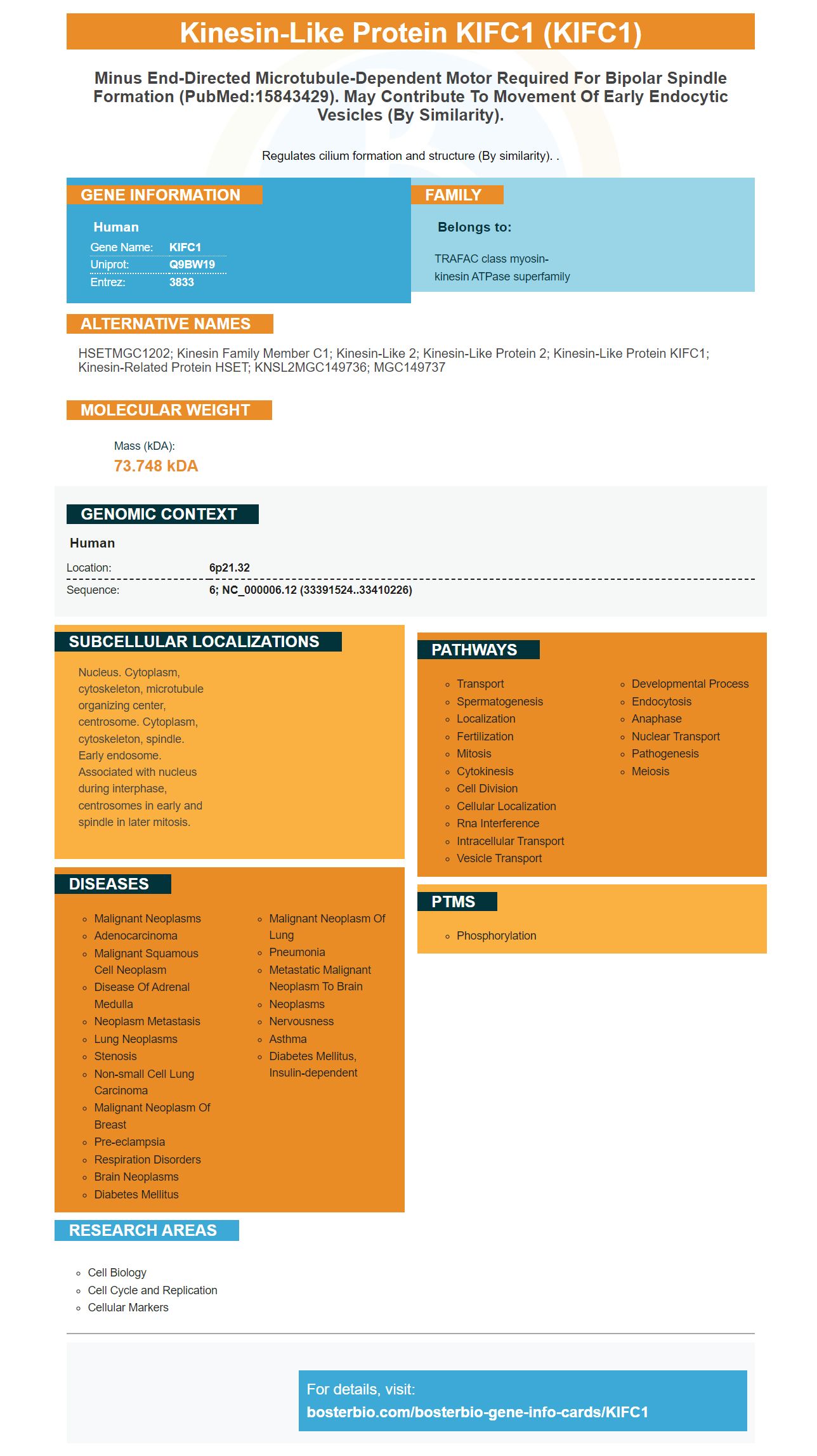

Facts about Kinesin-like protein KIFC1.

Regulates cilium formation and structure (By similarity). .
| Human | |
|---|---|
| Gene Name: | KIFC1 |
| Uniprot: | Q9BW19 |
| Entrez: | 3833 |

| Belongs to: |
|---|
| TRAFAC class myosin-kinesin ATPase superfamily |

HSETMGC1202; kinesin family member C1; kinesin-like 2; Kinesin-like protein 2; kinesin-like protein KIFC1; Kinesin-related protein HSET; KNSL2MGC149736; MGC149737
Mass (kDA):
73.748 kDA

| Human | |
|---|---|
| Location: | 6p21.32 |
| Sequence: | 6; NC_000006.12 (33391524..33410226) |
Nucleus. Cytoplasm, cytoskeleton, microtubule organizing center, centrosome. Cytoplasm, cytoskeleton, spindle. Early endosome. Associated with nucleus during interphase, centrosomes in early and spindle in later mitosis.


KIFC1 is an essential bipolar mitotic motor protein. Inhibiting invasion and cell migration, it also induces apoptosis. But what are the best uses of this antibody? Let's find out. KIFC1 antibody is available from Boster Bio (catalog number A05325). Here are the best uses of KIFC1 marker:
KIFC1 is a minus-end-directed motor protein with several functions. It participates in cell division and may promote tumor malignancy through interaction with cyclins and induces drug resistance to tamoxifane and taxanes. It has been linked to metastasis in NSCL and triple-negative breast cancer. It is functional in normal cells, where it is required for organelle trafficking, spermiogenesis, and oocyte development and gestation.
The kifer-like motor kinase (KifC1) is a central component of the mitotic spindle, and is involved in tethering the centrosomes and MTs together. In a cell, KifC1 promotes the assembly of the ASPP family. It may also play a role in regulating the organization of the MT.
NOD has unique mechanochemical properties. It is thought to move chromosomes by association with plus ends of polymerizing MTs. While direct evidence of this mechanism is lacking, high resolution imaging of NOD-coated chromatin stretching events indicates that NOD exhibits MT-plus end-directed motility. In contrast, NOD324CC does not exhibit this activity. Furthermore, NOD485CC has multiple regions predicted to be disordered. These regions center on proline and threonine residues at positions 430-480. These regions are similar to unconventional EB1-binding motifs in fungal species.
KIFC1 has a high expression level in human muscle and testis, indicating a potential role for the protein in cancer cells. HE staining of these two tissues reveals distinct histological properties. Seminoma cells lack the convoluted tubule found in the testis, while testis cells exhibit rounded, even-sized nuclei and are large. This transition from seminoma to muscle is indicative of the infiltration of cancer cells into the tissue.
KIFC1 is a member of the kinesin-14 family and contains a motor domain on its C-terminus. KIFC1 is implicated in many cancer types, but its role in testis cancer remains unclear. It is known to play a role in the handling of supernumerary centrosomes, and has a role in cell migration and chromosome instability.
KIFC1 is required for the formation of bipolar spindle and for maintaining the genomic stability of primary human fibroblasts. This protein also binds to ubiquitin and functions as a minus end directed motor, localizing on mitotic spindles and moving toward the poles of the spindle. KIFC1 is required for the formation of bipolar mitotic spindles and tethering centrosomes and MTs together.
In transgenic mice, knockout mice lacking KIFC1 did not exhibit gross mitotic spindle defects. This result suggests that Kif18 may have a role in the formation of the mitotic spindle, but its involvement is probably masked by redundant functions in other tissues. Other chromosome functions require Kif18, including congression, metaphase-to-anaphase transition, and spindle checkpoint.
In a previous study, a genetic mutation of the gene KIFC1 caused an asymmetric bipolar spindle, with monopolar spindles. KIFC1-deficient cells had monopolar spindles, whereas wild-type mice had asymmetric bipolar spindles and asymmetric bipolar division. Moreover, KO mice lacked KIFC1 exhibited asymmetric bipolar spindles. These cells also lacked Kif2A localization, which is essential for poleward MT flux.
In addition to regulating centrosome clustering, KIFC1 also controls the formation of unusual kinetochore attachments, which may help cancer cells achieve their characteristic bipolar spindle structure. In addition, it is a critical regulator of survivin in cancer cells. SAC complexes increase the rate of lagging chromosomes. This activity may enhance the malignancy of tumor cells.
KIFC1 is essential for bipolar cell division. Increasing expression of this protein in cancer cells increases cell survival and improves the likelihood of genetic mutation. Moreover, KIFC1 increases the chances of tumor malignancy. In addition, it promotes proper cell division and shortens the cell cycle. KIFC1 also contributes to tumor metastasis and cell proliferation.
Previous research has indicated that KIFC1 is required for meiosis I and meiosis II in oocytes. In addition, it plays an important role in centrosome formation in mouse oocytes. This suggests that it may also have a role in human oocyte meiosis. But further research is needed to fully understand the role of KIFC1 in bipolar mitosis.
MiR-1179, a mRNA target of PIK3CA, suppresses cell invasion and migration in A549 cells. It also inhibits the progression of glioblastoma cells through downregulation of the E2F transcription factor. MiR-1180 regulates p21(WAF1/CIP1) and promotes apoptosis in hepatocellular carcinoma cells via the NF-?B signaling pathway.
KIFC1 is a small RNA that suppresses the expression of several genes associated with tumorigenesis. It also inhibits the migration and invasion of glioblastoma cells, including MMP-14 and EZH2.
MiR-345 regulates non-small cell lung cancer cell migration and invasion. A group of researchers used two-hybrid assays to investigate its role in lung cancer. The study used 52 pairs of normal lung and NSCLC tissue samples to measure the NSCLC cells' migration and invasion capacities in vitro. Then, they used Western blot and qRT-PCR to determine the relative expression of YAP1 (yes-associated protein), a tumor-related gene.
In another study, KIFC1 siRNA blocked cell migration and invasion. When compared to a negative control group, KIFC1 siRNA significantly inhibited both cell migration and invasion. Moreover, this mRNA has a unique ability to suppress EMT, which is a hallmark of breast cancer. Further, it also inhibits the migration of breast cancer cells and osteosarcoma.
Inhibition of miR-10b through exosome-mediated transfer inhibits colorectal cancer metastasis by suppressing the expression of HOXD10. In addition, miR-10b also restricts the growth of breast cancer cells and promotes the transition from epithelial to mesenchymal stage. In addition, miR-10b inhibits cell invasion and migration in colon cancer cells via the Smad3-mediated tumor glycolysis.
A novel compound, KIFC1, inhibits gastric cancer cell migration and invasion. It targets the PPP1CA-Rb pathway, inhibiting the expression of SOX2, and suppresses growth and invasion. It also inhibits the production of mRNAs involved in gastric cancer. And microRNA-126 inhibits the expression of several genes that promote cancer. It is known to affect tumor cell migration and invasion, which are essential for gastric cancer metastasis.
The role of KIFC1 in regulating immune cells and tumor progression remains to be determined. Multiple genetic mutations in tumors increased their incidence, and the tumor microenvironment affects both cancer progression and the immune response. In recent years, immune checkpoint blockade (ICB) therapy has been developed to treat different types of cancer. However, biomarkers of response to ICB therapy may help to select which patients will benefit from this treatment.
KIFC1 regulates the activity of transcription factors that promote cell survival and differentiation. The protein interacts with the plus ends of microtubules, enabling them to slide antiparallel. The process leads to the formation of a pseudo-bipolar spindle, with lagging chromosomes. This may prevent premalignant cells from arresting mitosis.
KIFC1 expression correlates with tumor immune status. It is also associated with the expression of immune-related genes and immuno-checkpoints. In addition, KIFC1 expression is positively related to the progression of LIHC. Further, the KIFC1 gene level is associated with the expression level of immune-related genes and multiple cancer markers, including the inflammatory response and the tumor's immune status.
DTX-resistant PCa cells were treated with CW069, an allosteric inhibitor of KIFC1. CW069 treatment suppressed cell viability in DTX-resistant cell lines. Moreover, the drug's IC50 value was lower in DTX-resistant cell lines than in parental cells. Thus, DTX-resistant PCa cells may be a viable therapy for patients suffering from apoptosis-associated toxicity.
In previous studies, high KIFC1 expression was associated with poorer OS, which suggested that high KIFC1 expression may act as an independent biomarker for African-American triple-negative breast cancer patients. Moreover, KIFC1 differentially expresses in cytoplasm and nucleus, indicating that it may contribute to the progression of breast cancer. In addition, higher expression of KIFC1 in the nucleus may be associated with an increased risk of developing LIHC.
LIHC tissues were isolated from 37 patients with LIHC. Interestingly, KIFC1 expression was higher in LIHC tissue than in normal liver tissues. In addition, protein expression analysis indicated that KIFC1 is expressed near the nucleus of LIHC tumor cells, which contrasted with normal liver tissues. Furthermore, KIFC1 was not present in the cytoplasm of normal liver tissues.
PMID: 10369922 by Janitz K., et al. Genomic organization of the HSET locus and the possible association of HLA-linked genes with immotile cilia syndrome (ICS).
PMID: 8276466 by Ando A., et al. Cloning of a new kinesin-related gene located at the centromeric end of the human MHC region.


