This website uses cookies to ensure you get the best experience on our website.
- Table of Contents
1 Citations 5 Q&As
Facts about Potassium voltage-gated channel subfamily A member 1.
The station alternates between opened and closed conformations in response to the voltage difference across the membrane (PubMed:19912772). Can form practical homotetrameric channels and heterotetrameric channels that contain varying proportions of KCNA1, KCNA2, KCNA4, KCNA5, KCNA6, KCNA7, and possibly other relatives as well; station properties are based on the sort of alpha subunits that are part of the station (PubMed:12077175, PubMed:17156368).
Human | |
---|---|
Gene Name: | KCNA1 |
Uniprot: | Q09470 |
Entrez: | 3736 |
Belongs to: |
---|
potassium channel family |
AEMK; EA1; HBK1; HUK1; Kv1.1; MBK1; MGC126782; MGC138385; MK1; potassium voltage-gated channel subfamily A member 1; potassium voltage-gated channel, shaker-related subfamily, member 1 (episodicataxia with myokymia); RBK1; Voltage-gated K(+) channel HuKI; Voltage-gated potassium channel HBK1; Voltage-gated potassium channel subunit Kv1.1
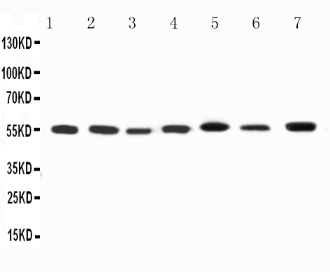
Mass (kDA):
56.466 kDA
Human | |
---|---|
Location: | 12p13.32 |
Sequence: | 12; NC_000012.12 (4909905..4918256) |
Detected adjacent to nodes of Ranvier in juxtaparanodal zones in spinal cord nerve fibers, but also in paranodal regions in some myelinated spinal cord axons (at protein level) (PubMed:11086297). Detected in the islet of Langerhans (PubMed:21483673).
Cell membrane; Multi-pass membrane protein. Membrane. Cell projection, axon. Cytoplasmic vesicle. Perikaryon. Endoplasmic reticulum. Cell projection, dendrite. Cell junction. Cell junction, synapse. Cell junction, synapse, presynaptic cell membrane. Cell junction, synapse, presynapse. Homotetrameric KCNA1 is primarily located in the endoplasmic reticulum. Interaction with KCNA2 and KCNAB2 or with KCNA4 and KCNAB2 promotes expression at the cell membrane (By similarity).
Boster Bio Anti-Kv1.1 potassium channel/KCNA1 marker is a high quality antibody that targets the KCNA1 protein. This article will describe what this antibody can do and the various platforms Boster uses to validate its products. Boster Bio is committed to developing high-quality antibodies and is committed to ensuring that they work well for your research.
This boster bio anti-Kv1.1 potassium channel/KCNA1.antibody is a part of their Picoband(tm) line. They have been testing this antibody using Western blotting and flow cytometry. Boster bio anti KCNA1 antibodies work with cell lysates from Rat, Mouse, and Human. Using the correct antibody will allow you to compare the levels of protein in human and mouse samples.
The brain, kidney, CNS, and brain all contain the KCNA1 protein. It forms heterotetrameric as well as homotetrameric potassium channels in membranes. The potassium channel regulates cell functions and controls neuronal excitability. Epilepsy, a condition characterised by frequent seizures, is associated with mutations in this genetic gene. The Boster Bio Anti-Kv1.1 potassium channel/KCNA1 antibody can help researchers identify the protein responsible for seizures in patients.
The protein mediates transmembrane potassium ions transport through excitable membranes. When voltage changes across the membrane, the potassium channel protein assumes a closed or open conformation, forming a potassium-selective channel. Its alpha and beta subunits bind KCNAB2 & PDZ domains of DLG1 & DLG2.
WB, ICC, or Immunofluorescence techniques are commonly used to identify target Antigens. ICC staining differs from IHC, which requires tissue preservation. ICC staining involves shorter fixation periods. Both types need antigen retrieval procedures. Both heat-induced epitope retrieval as well as proteolytic enzymatic retrieve can be used to retrieve epitopes that have been identified by antigenic targets.
Although WB is an excellent tool to validate antibodies, it is not the only option. While knockout animals are often the best method for antibody validation they should be used with caution. To avoid this, be sure to use Boster antibodies that are validated for use in ICC. Here are a few reasons why WB is the preferred method of antibody validation.
- The sensitivity of the MAP3K5 antibody was tested. It was successfully used for IHC to detect the protein from lung cancer tissue. The anti–ASK1/MAP3K5 is valid for WB. It can also be used in IHC, ICC. Immunofluorescence. Its sensitivities have been increased to the picogram scale, and the validation images can readily be obtained upon request. Supervision saves 30 minutes IHC and dramatically improves background contrast. - Picoband is powered by insights into the best immunogen design and manufacturing process. Picoband also offers technical support to support your success.
Monoclonal antibody. Monoclonal antibodies are antibodies that have been derived from a single cell of the B cell. They recognize an epitope differently. This type is only possible with a specific protocol. Monoclonal antibody reacts only with one epitope. The immune response of alpacas is weak compared to that of humans.
- IF staining: In this procedure, extracellular matrix is removed before performing the ICC or IF staining. IF staining, unlike WB and ICC, requires the removal of extracellular mater. In contrast, WB and ICC staining use cultured cells, IF is more sensitive. It uses more fluorescence to visualize the presence specific protein antigens.
Monoclonal anti CTLA4 antibody: This antibody recognizes CTLA-4 from human tissue. It has been tested for WB and IHC, ICC, as well as Immunofluorescence. This antibody was tested against more than 250 cell lines, both transfected and from tissue. It has a higher affinity than CD28 for ligands, but it is less expressed.
Monoclonal antibodies: IGY polyclonal antibodies are produced by 19 leading antibody providers. Monoclonal antibodies are more stable than polyclonal antibodies and can withstand repeated freeze/thaw cycles. Monoclonal antibodies can be used in immunotherapy and research. Antibodies are used to detect antigens in tumor cells.
The anti-Versican/VCAN antibody, part of the Picoband(tm) catalog, has been thoroughly validated in flow cytometry. The antibody reacts with FACS-confirmed human, mouse and Rat cell lines. Boster Bio validates all of its antibodies using ICC to ensure that they are both highly specific and highly sensitive. The manufacturer has a long history of supplying high-quality antibodies to laboratories worldwide.
PMID: 19912772 by Ramaswami M., et al. Human potassium channel genes: molecular cloning and functional expression.
PMID: 2128063 by Freeman S.N., et al. Cloning and characterization of a cDNA encoding a human brain potassium channel.
*More publications can be found for each product on its corresponding product page