This website uses cookies to ensure you get the best experience on our website.
- Table of Contents
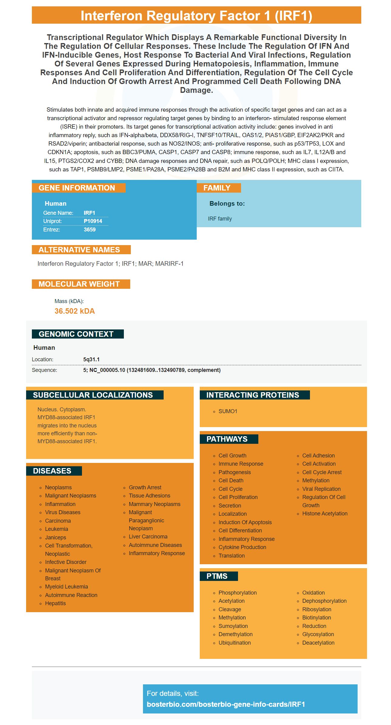
Facts about Interferon regulatory factor 1.
Stimulates both innate and acquired immune responses through the activation of specific target genes and can act as a transcriptional activator and repressor regulating target genes by binding to an interferon- stimulated response element (ISRE) in their promoters. Its target genes for transcriptional activation activity include: genes involved in anti inflammatory reply, such as IFN-alpha/beta, DDX58/RIG-I, TNFSF10/TRAIL, OAS1/2, PIAS1/GBP, EIF2AK2/PKR and RSAD2/viperin; antibacterial response, such as NOS2/INOS; anti- proliferative response, such as p53/TP53, LOX and CDKN1A; apoptosis, such as BBC3/PUMA, CASP1, CASP7 and CASP8; immune response, such as IL7, IL12A/B and IL15, PTGS2/COX2 and CYBB; DNA damage responses and DNA repair, such as POLQ/POLH; MHC class I expression, such as TAP1, PSMB9/LMP2, PSME1/PA28A, PSME2/PA28B and B2M and MHC class II expression, such as CIITA.
Human | |
---|---|
Gene Name: | IRF1 |
Uniprot: | P10914 |
Entrez: | 3659 |
Belongs to: |
---|
IRF family |
interferon regulatory factor 1; IRF1; MAR; MARIRF-1
Mass (kDA):
36.502 kDA
Human | |
---|---|
Location: | 5q31.1 |
Sequence: | 5; NC_000005.10 (132481609..132490789, complement) |
Nucleus. Cytoplasm. MYD88-associated IRF1 migrates into the nucleus more efficiently than non-MYD88-associated IRF1.
This article will focus on IRFs and how they can be used by the body. They play a variety roles that include tumor suppressive properties and regulate the immune system. They are also found in many cells and tissues including fibroblasts. This article will discuss the most efficient uses for this gene marker. Before we begin, let's go over some fundamentals. This gene is present in many tissues and cells like fibrocytes that make collagen and elasticin.
Many viruses target IRFs to evade the body's immune system. These proteins have important roles in managing immune responses, and they have significant tumor suppressive properties. Numerous viral factors have been discovered that target the IRF1 marker. These discoveries could result in more effective treatments for all forms of myeloid leukemia. In addition to targeting IRF1, Boster Bio's newest test could help in determining the best treatments for patients with this disease.
Polysaccharides, specifically those that are sugars can also act as in innate immune cells activators. They have been found to increase the immunogenicity of antigens. While further research is required to confirm this, the findings of Boster Bio's study suggest polysaccharides may enhance the immune system. Researchers who are looking for new ways to prevent and treat infections will be delighted to learn this.
IRF1 is responsible for the development of T cell responses as well as its immunomodulatory function. The T-cell receptors which activate and recognize T cells can influence the T-cell response. This protein is found in every hemocyte and regulates the immune response. These markers are essential for developing vaccines and drugs. It is important to comprehend their purpose.
The IRF1 marker also has been linked to a better understanding of how cancer cells react to antigens. These molecules can regulate T-cell responses to toxic substances. The IRF1 protein is found on the surface of the majority of human T-cells and is recognized by the Toll-like receptor 2/1 heterodimer on plasmacytoid DC. These molecules activate the PI3K pathway, and inhibit transcription factor GSK3b. They can also enhance the expression of anti-inflammatory genes via the CREB/CBP pathway.
Multivalent vaccines offer a way to boost the immune response. The vaccines contain various molecules that act on different receptors. They are able to interact more effectively in regulating the immune system. These vaccines are also cost effective and are suitable for developing countries. In addition to these vaccines, Boster Bio has also created a variety of other products. One of these products is the IRF1 vaccine. It was recently developed and licensed by Boster Bio.
IRF1 is an expression-related gene that is expressed in a variety of human cancers such as the ovarian, breast, and lung. It is believed to play a role in tumor-suppressive functions and could be a key factor in CRC metastasis. It is not clear what the role IRF1 is playing in CRC. In the present study, we demonstrate that the gene is expressed over in CRC tissue , but is not expressed in cancer cell lines.
We have identified 36 DEGs that are associated with inflammatory and immune responses, including T receptor signaling pathways and the differentiation of T helper 1 cells and CD8+ T-cells. We also identified four DEGs which significantly correlated with the stage of tumor and distant metastasis. Additionally, we found that upregulation of IRF1 was associated with a decrease in tumor size and distant metastasis suggesting the role of IRF1 in the regulation of immune evasion.
We also discovered that IRF1 loss affected the differentiation of NK cells and increased susceptibility to viral infections. In mice, loss of IRF3 also resulted in a decrease in the IL-6 response and the IL-12 response. Finally, in human breast cancer, loss of IRF7 results in metastatic growthand a deficient expression of IRF8 is a key factor in the Th2 response and differentiation of macrophages. It is also linked to the increase in proinflammatory cytokine levels as well as tumor cell survival.
It was discovered that IRF1 is involved in the regulation of gene expression and is found in a variety of gene promoter regions. We discovered that IRF1 can bind to the promoter region of the RASSF5 Gene in the course of a study. We also found that anti-IRF1-related groups were more enriched in the promoter sequence of RASSF5. These data suggest that IRF1 has an important role in the regulation of gene expression.
IRF1 is also linked to STAT1. Independent prognostic factors have been linked to breast cancer. Intense levels of STAT1 stimulation are associated with a longer duration of life. Additionally, STAT1 activation is associated with decreased cancer growth and an increase in apoptosis. Furthermore, STAT1 is known to suppress tumors in mouse models. Klover et al. Klover et al. found that mice deficient in Stat1 had a higher rate of mammary tumors growing than wild-type mice.
IRF1 is a transcription factor that regulates cardiac remodeling. Its deficiency can reduce cardiac fibrosis and hypertrophy. This means that it could represent a promising therapeutic target for cardiac remodeling. It isn't known at this time what diseases are associated with IRF1 expression. Here are a few conditions in which IRF1 is expressed:
The gene IRF1 is both transcriptional activator (or a transcriptional regulator) However, only a few studies have examined its non-coding RNA-related regulatory capabilities. The current study has identified miRNA-associated SNPs in the 3'UTR of IRF1. The study will investigate the functional relationship between IRF1 (and miRNAs) and its diagnostic utility. IRF1 plays a vital role in the control and maintenance of different cell types.
Down syndrome causes an increased amount of IFN receptors on 21 chromosomes. This causes a type 1 interferon gene signature. This means that IRF1 is constantly activated in fibroblasts. The increased risk of developing erosive or inflammatory arthritis has been due to the continuous activation and activation of interferon pathways. Further research is needed in order to find out if the genes found in IRF1 are directly involved in these conditions.
Studies have shown that IRF1 depletion may enhance the effectiveness of anti-VEGF therapy in the uterine leiomyosarcoma tumorigenesis. Additionally, it decreased the cytotoxicity of Dp in breast cancer cells. These findings suggest that IRF1 might play a crucial role in the formation of tumors. What is IRF1 exactly? How does IRF1 regulate the growth of tumors?
There are various types of IRF1 markers that are present in human fibroblasts. IRF1c is one of them. It is a transcriptional target of FOXM1c. The protein binds to the promoter region of IRF1 and is associated with cell migration and invasion. Patients with oesophageal carcinoma who have high levels FOXM1c and IRF1c have low survival rates and poor prognosis.
We don't have a clear understanding of the mechanisms that regulate IRF1 expression in fibroblasts. However the promoter methylation of the gene can alter the induction process of IRF1 by activated T cells. Interferons can be increased through blocking IRF1. Inducing IRF1 within fibroblasts can increase the risk of breast cancer.
Although IRF1 is expressed in a variety of different tissues it has been proven that it inhibits the function of NFkB and BCL2. It is unclear whether interferon g could help to resensitize breast cancer cells which are not able to respond to treatment with antiestrogens, despite these findings. Furthermore, the Schwartz-Roberts group recently showed that IRF1 is linked to autophagy in human breast tumors. ATG7 heterozygous mice are affected by the elevated levels of IRF1 in DMBA-induced breast cancers. In 107 matched human breast cancer tumors, nuclear IRF1 expression is in inverse correlation with ATG7 levels of the protein.
Recent studies have revealed that the inactivation of IRF1 alters transcriptional responses in the colon. The mutants lacking IRF1 had a higher infiltration of CD45+ cells at D26 compared to B6 controls. Three times more CD45+ cells were observed in Irf1colons / mutant colons than wild-type controls. The majority of Irf1cells found in the infiltrate were myeloid cells. The patients also had higher levels of eosinophils after they were carrying the IRF1 gene.
IRF1 proteins are genetically and structurally identical to other members of the transcription factor family. Their purpose is to regulate translocation. They also are able to bind to IRE elements found in promoters of the target gene. IRF proteins also play a role transactivation. They also inhibit NF kBa. Furthermore, since IRFs play a role in transactivation, they are especially important in the process of overcoming HIV infection.
The colon is just one of many tissues that have the IRF1 gene. It is yet to be understood exactly why IRF1 is expressed in these different tissues. Recent research has revealed that IRF1 plays a key function in colon tumor development However, the mechanisms behind it remain elusive. Interferon-g treatment causes an increase in IRF1 expression in RAW264.7 cells. In M1 macrophages miR-130b-3p is associated with IRF1 negatively.
IRF1 has been linked to CA-CRC. IRF1 loss can impact the differentiation of CD8+ cells. Irf1 knockout mice are less likely to differentiate into CD8+ T cells and NK cells in lung mononuclear cells. A tumor-permissible microenvironment is possible due to the disappearance of IRF1.
PMID: 2726461 by Maruyama M., et al. Sequence of a cDNA coding for human IRF-1.
PMID: 3409321 by Miyamoto M., et al. Regulated expression of a gene encoding a nuclear factor, IRF-1, that specifically binds to IFN-beta gene regulatory elements.