This website uses cookies to ensure you get the best experience on our website.
- Table of Contents
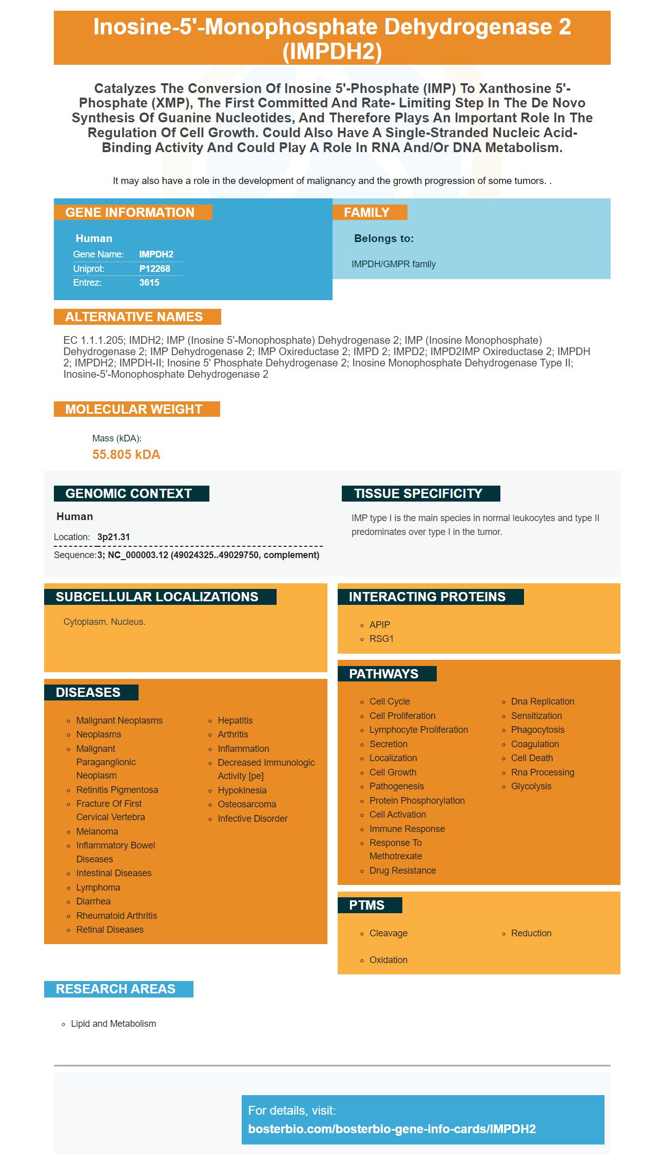
Facts about Inosine-5'-monophosphate dehydrogenase 2.
It may also have a role in the development of malignancy and the growth progression of some tumors. .
Human | |
---|---|
Gene Name: | IMPDH2 |
Uniprot: | P12268 |
Entrez: | 3615 |
Belongs to: |
---|
IMPDH/GMPR family |
EC 1.1.1.205; IMDH2; IMP (inosine 5'-monophosphate) dehydrogenase 2; IMP (inosine monophosphate) dehydrogenase 2; IMP Dehydrogenase 2; IMP Oxireductase 2; IMPD 2; IMPD2; IMPD2IMP oxireductase 2; IMPDH 2; IMPDH2; IMPDH-II; inosine 5' phosphate dehydrogenase 2; inosine monophosphate dehydrogenase type II; inosine-5'-monophosphate dehydrogenase 2
Mass (kDA):
55.805 kDA
Human | |
---|---|
Location: | 3p21.31 |
Sequence: | 3; NC_000003.12 (49024325..49029750, complement) |
IMP type I is the main species in normal leukocytes and type II predominates over type I in the tumor.
Cytoplasm. Nucleus.
This article provides a brief overview of the role of IMPDh2 and IMPDH2 in the EMT process and in cancer metastasis. It also briefly discusses the roles of these proteins in regulating intracellular GTP pools, and inhibiting the assembly of cytoophidia. It ends with recommendations for future research. Download Boster’s free IMPDH2 ELISA and Western Blotting assays.
In a recent study, we found that patients with ccRCC with the IMPDh2 mutation were more likely to have poor prognosis. This is because IMPDH2 increases the EMT process and decreases Ecadherin expression. IMPDH2 promotes CRC cell proliferation through activation of PI3K/AKT/mTOR, and FOXO1 signaling pathways.
CRC tumor cells showed a correlation between MET expression and IMPDh2. Interestingly, IMPDh2 regulates EMT signaling pathways, but its role in tumor progression is still not fully understood. This gene is a potential cancer-causing oncogene that could potentially be used as a prognostic marker. However, further studies are required to validate the relationship between IMPDh2 and EMT.
In a previous study, we discovered that IMPDh2 regulates tumor metastatics via EMT signaling pathways. This gene also regulates cell morphology in malignant and normal cells, promoting tumor growth and invasiveness. It is important to remember that IMPDh2 expression strongly correlates with the presence other EMT markers like p21Cip1 (or p27Kip1).
In addition to regulating tumor growth, IMPDh2 also regulates cell proliferation and metastasis through the PI3K/Akt and FOXO1 pathway. This discovery is a landmark for cancer research. This protein is a key regulator to EMT and can potentially be used therapeutically. It may also play an important role in tumor growth. In this instance, IMPDh2 inhibition could have a therapeutic advantage.
EMT is a tightly controlled process. Tumor cells that undergo EMT acquire the characteristics of mesenchymal cells, which increases their metastatic potential. This metastatic ability allows epithelial-cancer cells to enter and grow in different tissues. This requires constant nourishment and metabolic reprogramming. Thus, inhibiting EMT might be an attractive treatment strategy.
IMPDh2 plays a vital role in EMTTFs. It inhibits the major PFK1 isoform and promotes the production PPP in breast cancer cell line lines. PPP generates NADPH, a reducing equivalent, and precursors for the synthesis of amino acids, fatty acids, and nucleotides. EMT is also inhibited in cell lines that have PGI. However, it is necessary to conduct a more thorough analysis to confirm its role as a tumor-growth and metastasis inhibitor.
The progression and development of the cell cicle is directly related to the expression IMPDh2 in human lymphocytes. CRC cells with high levels of IMPDH2 have a higher proportion in the S phase and a lower G0/G1 stage. Silencing IMPDH2 reduces the number in the S-phase and increases G0/G1 levels.
ccRCC tumor metastasis is associated with IMPDh2-assembled cytoophidia. Moreover, YB-1 proteins are translocated into the cell nucleus following interaction with IMPDh2. MPA-induced cytoophidia raises YB-1 expression, which correlates with tumor metastasis. So, it seems that the expression of IMPDh2 is critical for tumor metastasis.
IMPDH2 regulates intracellular GTP levels. This enzyme has a dual role as a GTPase, regulating the activity of RAC1 and the GTPase-like protein (IMPDH2). Cell cultures show filament formation when intracellular glutamate levels decrease. Fibril formation is thought to promote cell proliferation and enzymatic activity. Recent research has investigated how filament formation is controlled. Fernandez -Justel et. al. Fernandez et al. showed that filament formation was more resistant to allosteric inhibition.
A mutation of IMPDH or HPRT can cause a change in purine nucleotide levels. The intracellular GTP levels in splenocytes were measured during resting conditions and mitogen stimulation. GTP levels in IMPDH2-mutant mice was 30% lower than that of wild-type mice. GTP levels slightly decreased when cells were stimulated using Con A. However, this did NOT reach statistical significance. Moreover, the levels of ATP were not significantly different between wild-type and mutant mice.
IMPDH2 plays a significant role in regulating the intracellular TTP pool. It is crucial for the synthesis RNA and DNA. It also plays a role in signaling transductions. De novo GTP, AMP biosynthesis pathways require two ATPs each and three reactions, while salvage synthesis requires nine ATPs for twenty reactions. Both pathways share a common metabolic pathway that converts glucose to IMP, which is GTP's substrate.
Although the protein level for IMPDH2 is nearly identical in humans, there are additional mechanisms to regulate its activity. Although it is known that PI-3 kinase activates IMPDH2 function, it is not yet clear which sites are phosphorylated via PKB/AKT. It is also not known what the exact role is of phosphorylation. There are multiple overlapping phosphorylation site on IMPDh2, but we don't know what those domains are.
Increasing levels GMPR activities trigger filamentation IMPDH. GMPR overexpression, however, does not influence intracellular GTP levels. Inhibition of IMPDH2 activity does not alter the proliferation rate of IMPDH cells. CTP levels are increased, which promotes IMPDH cytoophidium construction. These results support the role played by IMPDH in cellular signaling.
Guanine nucleotides regulate both IMPDh2 (and IMPDH2). Mutations affecting both genes disrupt this allosteric inhibition. Some mutations in IMPDH2 can lead to retinopathies. These mutations can alter intracellular GTP levels. Normal cell growth depends on the IMPDH2 gene located in the early neural crest.
A recent study found that mice lacking IMPDH2 were unable to differentiate cytotoxic T lymphocytes. GTP levels were up 7.7-fold (+2.6%) in IMPDH II/ mice when Ab stimulation was applied. Despite the fact that the mice did not grow normally, their spleens did respond to anti-CD3 and anti-CD28 stimulation.
The ability to assemble cytoophidias is a conserved phenomenon that has existed throughout the history of evolution. CTPS-cytoophidia were first found in fruitflies and later in bacteria, yeast, mammalian and mammalian spores. IMPDH cytoophidia are only found in mammalian cell lines. IMPDH cytoophidia have been observed in some cell types in normal conditions. However, they are absent in cells treated using glutamine analogues and deazauridine. The cells can also form filaments from many metabolic enzymes.
In worms, IMPDh2 & IMPDH2 expression decreases to near undetectable levels at the pronuclear and then resumes at 2-cell stages. GTP levels high in the cytoophidium make cytoophidia assembly stronger. GTP levels are also controlled through IMPDh2 & IMPDH2.
The IMPDh2 & IMPDH2 protein are expressed dynamically by granulosa oocytes. IMPDh2 & IMPDH2 both inhibit meiosis during oocyte development. Moreover, inhibition of IMPDH activity compromises meiotic competence in oocytes and preimplantation embryo development.
After six hours of MPA treatment in 25 mM NPPC, COCs resumed meiotic activities. However, IMPDh2 & IMPDH2 failed form cytoophidia. MPA induced GVBD with a dose-dependent manner. For example, COCs cultured in 100 mM MPA for 2 h formed 83.7% type I cytoophidia. However, COCs had a 53.1% GVBD rate.
Although this study indicates that IMPDh2 & IMPDH2 are capable of coassembling, the mechanisms underlying filament assembly are still not clear. IMPDH and CTPS share similar catalytic activity. However, the filament assembly mechanism could sequester IMPDh2 from noncatalytic functions to scaffold other proteins. The presence of separate-offunction mutants in IMPDH will help us to address the role of assembly in vivo.
IMPDh2 is an essential component of the rod and ring macrostructure in ovaries. IMPDH activity plays a crucial role in oocyte growth and maturation. The IMPDH protein is found in mouse ovaries. A study of IMPDh2 in mice has shown that this enzyme can prevent meiotic arrest.
IMPDH2 is found in the oocyte and preimplantation embryos. Inhibition by IMPDH2 causes GVBD to be delayed in cultured denuded or cumulus-enclosed follicles. However, inhibition of IMPDH2 did NOT affect the extrusion the first polar-body. These results suggest that IMPDH2 is responsible for oocyte aggregation and disrupts oocyte IMPDH2 activities.
The study's authors propose that light-dependent phosphorylation (IMPDh2) regulates GTP/GDP levels. This conclusion is based on their observations. However, the authors didn't specify which IMPDh2 was more phosphorylated at the retina. Researchers should not conclude that light-induced photophosphorylation inhibits cytoophidia assembly if they don't have such information.
PMID: 2902093 by Collart F.R., et al. Cloning and sequence analysis of the human and Chinese hamster inosine-5'-monophosphate dehydrogenase cDNAs.
PMID: 1969416 by Natsumeda Y., et al. Two distinct cDNAs for human IMP dehydrogenase.