This website uses cookies to ensure you get the best experience on our website.
- Table of Contents
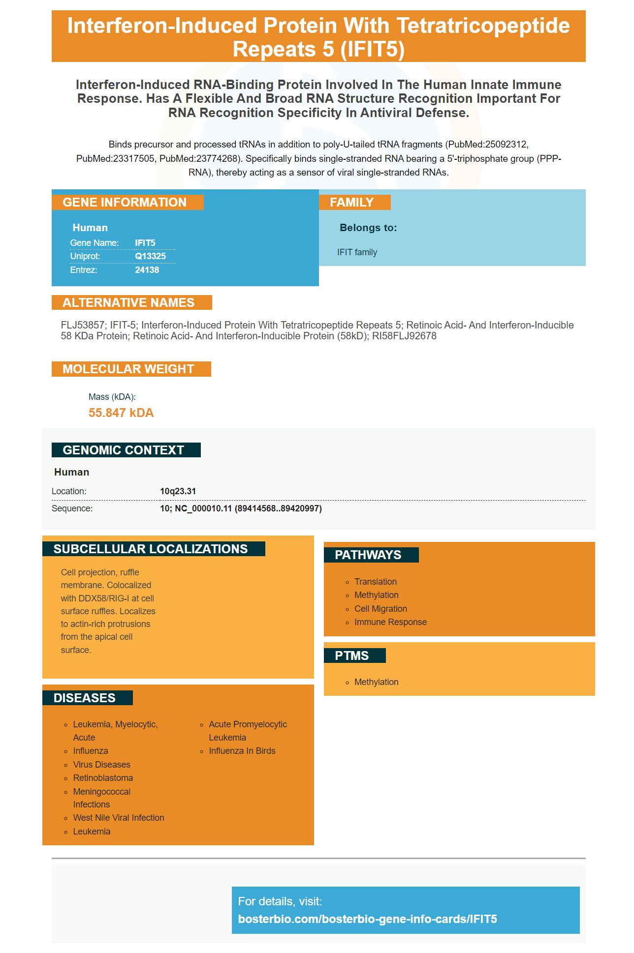
Facts about Interferon-induced protein with tetratricopeptide repeats 5.
Binds precursor and processed tRNAs in addition to poly-U-tailed tRNA fragments (PubMed:25092312, PubMed:23317505, PubMed:23774268). Specifically binds single-stranded RNA bearing a 5'-triphosphate group (PPP-RNA), thereby acting as a sensor of viral single-stranded RNAs.
Human | |
---|---|
Gene Name: | IFIT5 |
Uniprot: | Q13325 |
Entrez: | 24138 |
Belongs to: |
---|
IFIT family |
FLJ53857; IFIT-5; interferon-induced protein with tetratricopeptide repeats 5; Retinoic acid- and interferon-inducible 58 kDa protein; retinoic acid- and interferon-inducible protein (58kD); RI58FLJ92678
Mass (kDA):
55.847 kDA
Human | |
---|---|
Location: | 10q23.31 |
Sequence: | 10; NC_000010.11 (89414568..89420997) |
Cell projection, ruffle membrane. Colocalized with DDX58/RIG-I at cell surface ruffles. Localizes to actin-rich protrusions from the apical cell surface.
Boster Bio scientists might already be familiar with the IFIT5 markers' benefits and advantages. It allows you to submit the results of your experiments for species, applications, and special samples. It also allows you to receive product credits, which can be used by scientists all over the globe. The IFIT5 marker is the industry's first antibody probe, and its application is wide-ranging.
Boster Bio protein transfers by membrane staining are important factors in evaluating their effectiveness. If proteins are lost during transfer or the membranes are too thick, this can reduce the efficiency of the transfer. These problems can decrease the sensitivity and reproducibility of the transfer process. We have developed a variety improved fixative reagents and electrode materials to improve the efficiency of this method.
Ponceau S is the basis for Boster Bio's protein transfer by membrane staining. Ponceau S can be used to visualize the protein transfer. There are ready-to–use commercial stains available for this method. The signal is higher when the membrane is treated with alkali before staining. Proteins less than 2 ng per band will be visualized as red bands on pink backgrounds. The concentration of the gel is important as the higher the gel concentration, the slower the transfer efficiency.
For this test, we used a mixture of pooled human serum samples and 10% SDS-PAGE. The proteins were then transferred onto PVDF or Nitrocellulose membranes for further fixation treatments. We used biotinylated lectins by Vector Laboratories (Burlingame), CA, USA, Proteintech Group (Chicago, IL, USA) and Boster Corporation (Wuhan, China).
To perform this test, we first need to identify which proteins are present in the sample. We can then determine the amount of protein in the sample by using the appropriate antibodies for each protein. It is important to use the correct antibody for each protein in order to determine the efficiency and effectiveness of membrane staining. Boster will send high-quality digital images to analyze the sample once it has been determined. Boster Bio membrane staining is a great way to enhance your research.
Coomassie Brilliant Blue is another popular way to stain proteins. This stain is easy to see and inexpensive. It binds to proteins through ionic or hydrophobic interactions. This stain is compatible to blood proteins and can be used in bioethics as well as forensics. These are just a few benefits of Boster Bio membrane staining for protein transfer efficiency.
Boster Bio's IFIT5 protein transfer efficiency by ECL chemical chemiluminescence detection system was created to increase the efficiency of proteins in a wide variety of samples. The system is based upon antibodies that are conjugated to HP and emit strong blue light at around 450nm. The light emission takes place during the enzyme-substrate reactions and stops when the substrate is exhausted.
Optimal western blotting conditions are critical for obtaining accurate, reproducible results. The blot should then be exposed to optimal conditions of chemiluminescence for six to twenty hours to achieve the best results. The enzyme-to substrate ratio and the substrate concentration will determine the duration of light generation. While a blot shows a steady amount of enzyme, too much antibody-conjugate combination can cause a variable signal and low sensitivity. Boster Bio’s IFIT5 technology ensures that each component of protein transfer is optimized by its sensitivity and signal emission.
The laboratory uses the IFIT5 marker to test for viral RNA. The protein includes 482 amino compounds and eight TPR repeateds. It is a member IFIT and localizes in the Cell projection. It is a key component of virus detection in Molecular biology. Boster's antibodies are highly specific and are frequently cited in research. The antibodies are validated using Western Blotting, Immunohistochemistry, and ELISA.
Boster Bio's Western blotting system IFIT5 includes detection methods of Interferon-induced, 58-kDa protein. (RI58) is a 482-amino Acid protein that contains eight TPR repetitions. Located in the Cell projection, this protein functions as a sensor of viral RNA. Each antibody in this system is validated using a positive and a negative control.
To detect a target protein, the Western blotting process uses a gel containing the target protein. The gel's gel pattern is retained and is then probed using primary antibodies. Secondary antibodies are then used for the detection of the antigen. These secondary antibodies are usually labeled using a radioisotope or fluorophore. The intensity of a specific reaction gives information about the protein expression.
The Antibody Diluent Buffer should be used to dilute the primary antibody, preferably to 5%. The primary antibody should remain incubated for at minimum one hour and preferably overnight. To remove unbound antibody, the sample should be washed with TBS Wash Buffer three times. The secondary antibody can then be added to your blot. This should be repeated a second time.
A primary antibody is a must for performing a successful Western blot. There are many ways to detect the antibodies. One common method is polyacrylamide gel electrophoresis. Polyacrylamide gel electrodephoresis allows for the separation of proteins by molecularweight. This process can be hampered by a variety of factors. Protein degradation and glycosylation make a protein appear larger than predicted. Non-specific antibody binding, membrane drying, and other causes could also be responsible.
PMID: 9398535 by Niikura T., et al. A novel interferon-inducible gene expressed during myeloid differentiation.
PMID: 20950130 by Fensterl V., et al. The ISG56/IFIT1 gene family.