This website uses cookies to ensure you get the best experience on our website.
- Table of Contents
2 Citations 1 Q&As
2 Citations 6 Q&As
Facts about Indoleamine 2,3-dioxygenase 1.

Tryptophan shortage inhibits T lymphocytes division and accumulation of tryptophan catabolites induces T-cell apoptosis and differentiation of regulatory T-cells (PubMed:25691885). Acts as a suppressor of anti-tumor immunity (PubMed:23103127, PubMed:25157255, PubMed:14502282, PubMed:25691885).
| Human | |
|---|---|
| Gene Name: | IDO1 |
| Uniprot: | P14902 |
| Entrez: | 3620 |

| Belongs to: |
|---|
| indoleamine 2,3-dioxygenase family |

3dioxygenase; EC 1.13.11.52; IDO; IDO1; IDOIDO-1; INDO; INDOindole 2,3-dioxygenase; Indoleamine 2; indoleamine 2,3-dioxygenase 1; Indoleamine 2,3-dioxygenase; indoleamine-pyrrole 2,3 dioxygenase; Indoleamine-pyrrole 2,3-dioxygenase
Mass (kDA):
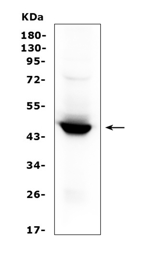
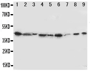
45.326 kDA

| Human | |
|---|---|
| Location: | 8p11.21 |
| Sequence: | 8; NC_000008.11 (39913891..39928790) |
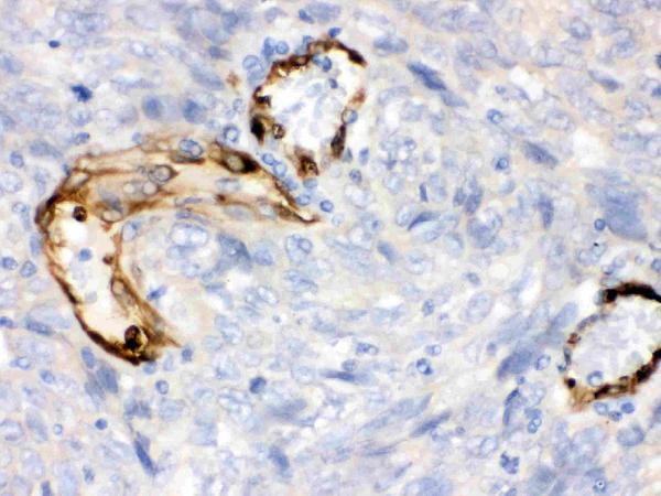
Expressed in mature dendritic cells located in lymphoid organs (including lymph nodes, spleen, tonsils, Peyers's patches, the gut lamina propria, and the thymic medulla), in some epithelial cells of the female genital tract, as well as in endothelial cells of term placenta and in lung parenchyma (PubMed:25691885). Weakly or not expressed in most normal tissues, but mostly inducible in most tissues (PubMed:25691885). Expressed in more than 50% of tumors, either by tumoral, stromal, or endothelial cells (expression in tumor is associated with a worse clinical outcome) (PubMed:18418598). Not overexpressed in tumor- draining lymph nodes (PubMed:26155395, PubMed:25691885).
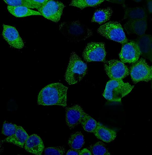
Cytoplasm, cytosol.


PMID: 2109605 by Dai W., et al. Molecular cloning, sequencing and expression of human interferon- gamma-inducible indoleamine 2,3-dioxygenase cDNA.
PMID: 2326172 by Tone S., et al. Primary structure of human indoleamine 2,3-dioxygenase deduced from the nucleotide sequence of its cDNA.
*More publications can be found for each product on its corresponding product page