This website uses cookies to ensure you get the best experience on our website.
- Table of Contents
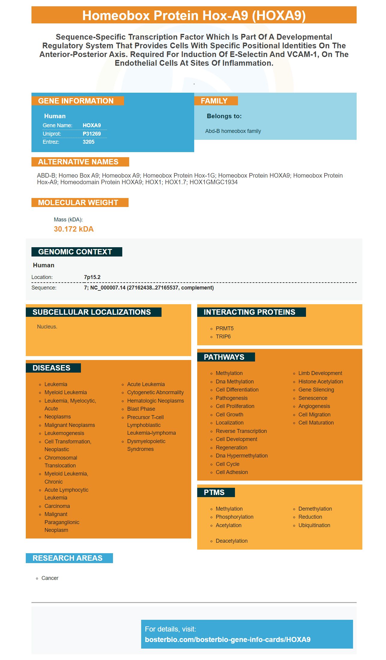
Facts about Homeobox protein Hox-A9.
.
Human | |
---|---|
Gene Name: | HOXA9 |
Uniprot: | P31269 |
Entrez: | 3205 |
Belongs to: |
---|
Abd-B homeobox family |
ABD-B; homeo box A9; homeobox A9; Homeobox protein Hox-1G; homeobox protein HOXA9; homeobox protein Hox-A9; homeodomain protein HOXA9; HOX1; HOX1.7; HOX1GMGC1934
Mass (kDA):
30.172 kDA
Human | |
---|---|
Location: | 7p15.2 |
Sequence: | 7; NC_000007.14 (27162438..27165537, complement) |
Nucleus.
This article will be of great help to anyone who is interested in satellite cell technology. This article will focus on the HOXA9 marker, and how it is used in satellite cell development. This article doesn't just cover these markers. Click the links below to learn more about them. You can also read more about MEIS1 (and PBX3).
The gene HOXA9 has been extensively used in gene therapy. This marker can be found in human leukemia cell lines. We used a recombinant HOXA9 human protein, which was made in E. coli. The His-Tag label was applied to the protein. It does NOT contain endotoxin like the original HOXA9 HOXA9 gene and was stored at -20 – 80 degrees Celsius.
HOX gene transcription factors regulate gene expression. They bind to DNA through the homeobox domain and exhibit high levels of redundancy in binding site specificity. They are found in heterodimeric and trimeric combinations and are often cofactors with a gene coding enzyme called HOX. These cofactors act in conjunction with the HOX gene, allowing it to activate and regulate target genes.
HOXA10 has been well-characterized as a marker of endometrial receptivity. It has pleiotropic effect on multiple aspects, including pinopode formation, stromal decidualization and leukocyte penetration, in adult endometrial growth. This gene regulates expression of multiple downstream targets, such as ITGB3, EMX2, HOXA9
The HOXA9 gene encodes a protein that is a member of the highly conserved HOX protein family. This gene is required for the differentiation and development of cells and plays important roles in hematopoiesis. It promotes proliferation, but it is downregulated during differentiation. The HOXA9 gene in acute myeloid leukemia is upregulated in almost 50% of cases. It is also accompanied by a cofactor.
The HOXA9 Gene and MEIS1 Gene are regulated by distinct pathways. These differences could reflect the diverse functional complexes that exist in leukemia cell lines. Huang et al. Huang et.al. These studies suggest that the HOXA9 mark is required for determining regulation of HIWI genes.
While there is no conclusive evidence to support the association between MEIS1 gene and ESCC risk (although the two genes are known to be associated with oncogenic activities), MEIS1 also correlates with stem cell markers. The MEIS1 genes is one of many genes involved in the development ESCC. Although MEIS1 has not been shown to be associated with ESCC risks factors, it may still be a good candidate for clinical trial.
The role of MEIS1 in cancer cell proliferation and differentiation has been suggested by studies involving mice. MEIS1 also plays a role in regulation of cellular metabolism, hematopoiesis, and other functions. MEIS1 could be involved in cancer therapies targeting cancer stem cell cells. These studies will allow clinicians and researchers to determine if MEIS1 plays a role in the creation of ESCC.
PBX3 is a great target for cancer research and also important in the treatment of many diseases such as leukemia or glioma. It is believed to play an oncogenic role in cancer, so targeting PBX3 would be extremely beneficial. To determine the most effective treatment, you must first identify the patient’s type of leukemia and HOXA9 status.
The HOXA9 antibody provides a reliable and reproducible method to measure the protein level of human cells. Boster's pipettes have high sensitivity and a wide range of dispensing capacities. Multichannel pipettes are best if you plan to use large samples. PBX3 Boster Bio: Best Uses Of The HOXA9 Marker
The HOXA9 gene is a hallmark of mixed lineage leukemia (MLL). This marker has been found to be overexpressed in a variety different cancers. PBX3 overexpression was sufficient to transform normal mouse stem cells. Despite this fact MEIS1's binding ability with PBX3 protein is critical for Hoxa9 and MLL-fusion proteins. Furthermore, overexpression of PBX3 is sufficient to induce leukemia.
Co-expression of PBX3 with MEIS1 recapitulates the core transcriptome of MLL-rearranged AML in mice. 160 genes were upregulated in co-expression of PBX3/MEIS1 and PBX3. But forced expression of HOXA9 or MEIS1 alone cannot achieve sufficient abundance of both markers. Ultimately, the best treatment is one that maximizes the expression of PBX3 and MEIS1 simultaneously.
To understand how satellite cells grow and differentiate, we investigated the role played by HOXA9. This protein is expressed in axons and controls satellite cell development. By blocking HOXA9 expression, we could determine whether satellite cells can differentiate into myotubes. Satellite cells have a limited capability for differentiation so HOXA9 control can be helpful in cell therapy.
Our experiments were conducted by transfecting rat HOXA9 coding segments into a pPLK/GFP plasmid. After cloning the rat HOXA9 coding regions was transfected into a pIRES2-DsRed2 virus. After transfection of primary satellite cells, they were divided into four different groups and incubated for 24 hours with Lipofectamine3000. After reaching confluence of sixty-seven percent, the cells were used in subsequent experiments.
After denervation HOXA9 expression shifted from nucleus to cytoplasm. It was detected in cells with a distorted nucleus. The expression of HOXA9 protein was also increased in satellite cells in the denervated groups. The cytoplasmic HOXA9 expression was significantly higher than that of the sham group.
The HOXA9 genetic was discovered in aged skeletal muscular tissue. This protein is highly active and restricted the development and function in satellite cells. This mechanism activates signaling pathway that are related to senescence. This has attracted considerable attention, especially for the treatment or prevention of aging. But, it is necessary to continue research in order to understand how satellite cells form and differentiate.
Fiber diameter analysis was carried out to confirm immunohistochemical results for HOXA9 Fiber diameter in the sham group increased with time, while in the denervated group it decreased. The distribution of fiber size shifted to the left, with a higher proportion of small fibres. A standard protocol was used for assessing fiber diameter. The results showed that immunohistochemical staining to HOXA9 is an effective method of measuring muscle fiber diameter.
Five candidate SNPs make up the TASP1 gene, and each one contributes to fiber diameter. The box plots show the median, 25th-75th, maximum, and minimum values as well as outliers. The HOXA9 marker was used for identification of the two candidate SNPs to TASP1 as being the most relevant for fiber diameter analysis. This marker was found in a study that examined genetic variance in muscle fiber diameter, a critical factor in determining the quality of wool.
The Pekin duck and mallard muscle fiber images were collected and fixed with 4% paraformaldehyde. Sections were stained using eosin and hematoxylin. Image-Pro Plus 6 software was used to measure the histomorphology and diameter of the fibers. Point-counting stereology was used as a method to estimate how many fibers were in each muscle fiber.
The study aimed to discover the mechanism that regulates HOXA9 in skeletal muscles. The HOXA9 gene was found to be highly active in older skeletal muscle and limits the function of satellite cells and muscle regeneration. This new system, which is focused on satellite cells, could be useful in the clinical treatment of deervated muscles. This could be a promising drug candidate for treating denervated muscle if further research is done.
PMID: 9880515 by Patel C.V., et al. Endothelial cells express a novel, tumor necrosis factor-alpha- regulated variant of HOXA9.
PMID: 8563754 by Borrow J., et al. The t(7;11)(p15;p15) translocation in acute myeloid leukaemia fuses the genes for nucleoporin NUP98 and class I homeoprotein HOXA9.