This website uses cookies to ensure you get the best experience on our website.
- Table of Contents
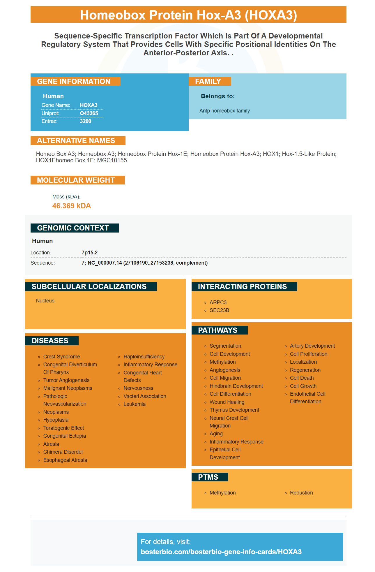
Facts about Homeobox protein Hox-A3.

| Human | |
|---|---|
| Gene Name: | HOXA3 |
| Uniprot: | O43365 |
| Entrez: | 3200 |

| Belongs to: |
|---|
| Antp homeobox family |

homeo box A3; homeobox A3; Homeobox protein Hox-1E; homeobox protein Hox-A3; HOX1; Hox-1.5-like protein; HOX1Ehomeo box 1E; MGC10155
Mass (kDA):
46.369 kDA

| Human | |
|---|---|
| Location: | 7p15.2 |
| Sequence: | 7; NC_000007.14 (27106190..27153238, complement) |
Nucleus.


If you are looking to find a reliable HOXA3-marker antibody, keep reading. Boster Bio has published its product review, which includes a comparison with Anti-Hoxa5. To learn more, read the review. Then, order your HOXA3 antibody to start your next experiment. We'll also cover the use and Anti-Hoxa5 antibody.
Boster Bio Anti-Homeobox A3 HOXA3 marker is designed to target homeobox mRNA. These proteins are known for controlling the expression of a variety of genes that regulate the growth of cancer cells. The HOXA3Marker is a single molecule antibody that detects mRNAs for HOXA3 (and HOXD10). Boster Bio Anti-Homeobox protein-A3 (HOXA3) mRNA comes in different sizes and can also be customized to suit your needs.
There are many suppliers who offer anti-HOXA5 antibodies that target the human protein and gene 'homeobox a5". These antibodies may also recognize homologs found in canines, monkeys, and porcine species. In some instances, the antibody may be used for diagnostic or therapeutic purposes. Continue reading to learn about the many benefits of HOXA5 –targeted reagents.
The researchers used a combination HOXA5/HOAIR antibodies to measure apoptosis in HL-60 HL-60 cells. This antibody inhibited cell death and increased the number HL-60 cells in G0/G1. The S phase had a significantly lower percentage for the group that received anti-HOXA5 plus sh-HotAIR.
Researchers used reverse transcription as well as western blot to determine HOXA5 protein expression. Knockdown HOXA5 reduced cell proliferation and led to apoptosis. Survivin expression was decreased and RNA-mediated apoptosis increased. Further research is necessary to determine the precise roles of HOXA5 within leukemia and in other diseases.
HOXA5 is a functional tool that can also suppress metastasis in lung cancers. This antibody blocks calcium-mediated polymerization actin. Furthermore, it suppresses cell proliferation and inhibits apoptosis, two essential processes for leukemia development. HOXA5 is expressed in leukemia cells and is associated with breast cancer, non-small cell lung cancer, and acute myeloidleukemia.
Boster bio also has an anti HOXA5 anti antibody. This antibody is very specific and can be used in research as well as clinical applications. The antibody recognizes HOXA5 as well as CYP-Dnmt3b. It can also be used to fight a variety other cancers and inflammatory disorders. Its use has opened doors to further research. This antibody can now be confidently used in your own research.
Primary antibodies were rabbit monoclonal and rabbit polyclonal Anti-HOXA5 respectively. They were diluted with 1 to 500 and 1 to 1000, respectively. The membranes had to be incubated overnight at 4°C. After incubation, the primary antibodies could be washed and then reacted with a sample. A loading control was provided by a rabbit polyclonal, anti-b-actin antibody.
The Meis1 gene is found in a subset perivascular fibroblasts, and pericytes in normal human health. It is a marker for myofibroblasts in aging. Meis1 antibody detects this marker in the HOXA3-tagged cells. Boster Bio first discovered Meis1 in 1997. It is one of most widely used markers to study aging.
Meis1 is expressed by mesenchymal kidney cells. Meis1 expression is found in interstitial myofibroblasts following injury. Meis1 antibody can detect mesenchymal medullary cells in the UUO, IRI regions. The Meis1 marker is capable of detecting mesenchymal tissues in a wide range human tissues, including the kidney.
The Meis1 MeRNA and Meis1 protein levels increased in elderly kidneys but did no change the severity renal fibrosis. In young, uninjured mice, the Meis1 protein was expressed in kidney fibrocytes. However, it was not expressed in injured or diseased mice. The expression of Meis1 was upregulated during differentiation and proliferation. Meis1 is not required for normal kidney development, unlike other kidney markers. A loss of Meis1 in the kidney had no effect on the severity of murine fibrosis.
The Meis1 protein is a member of the TALE family of homeobox-containing proteins. It binds Pbx proteins and Hox proteins. Its overexpression suggests that it may play a role in oncogenesis. Meis1 is related to the pre-B-cell leukemia transcription factor Mrg1.
This antibody is made from two different molecules: FCAMR and MEIS1. It uses the HOXA3 markers to recognize Meis1. These two antibodies are the only two available that use the HOXA3 marker. They are the only antibodies that use Meis1 for mouse models. They are not yet approved by FDA but are in the process.
Meis1 is an endogenous homeobox protein that is expressed in adult mouse kidneys and perivasculature. Meis1 antibodies stain nuclei and cytoplasm with eGFP. In adult mice, Meis1 is more abundant in the interstitial compartment than in the endothelium, and it is present around the blood vessels.
PMID: 12690205 by Scherer S.W., et al. Human chromosome 7: DNA sequence and biology.
PMID: 12853948 by Hillier L.W., et al. The DNA sequence of human chromosome 7.


