This website uses cookies to ensure you get the best experience on our website.
- Table of Contents
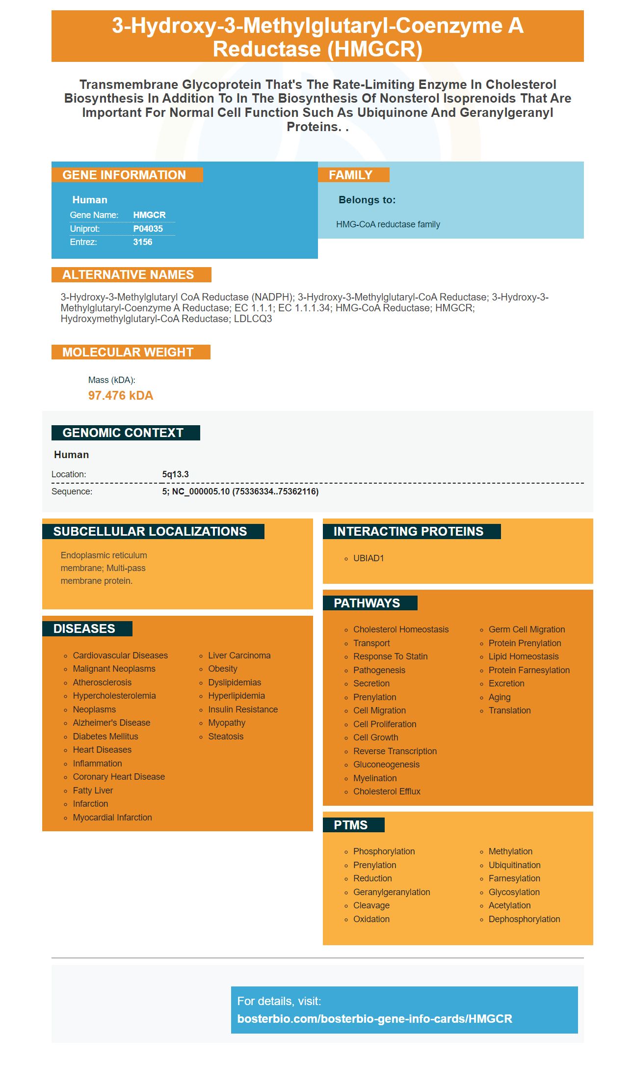
Facts about 3-hydroxy-3-methylglutaryl-coenzyme A reductase.
Human | |
---|---|
Gene Name: | HMGCR |
Uniprot: | P04035 |
Entrez: | 3156 |
Belongs to: |
---|
HMG-CoA reductase family |
3-hydroxy-3-methylglutaryl CoA reductase (NADPH); 3-hydroxy-3-methylglutaryl-CoA reductase; 3-hydroxy-3-methylglutaryl-Coenzyme A reductase; EC 1.1.1; EC 1.1.1.34; HMG-CoA Reductase; HMGCR; Hydroxymethylglutaryl-CoA Reductase; LDLCQ3
Mass (kDA):
97.476 kDA
Human | |
---|---|
Location: | 5q13.3 |
Sequence: | 5; NC_000005.10 (75336334..75362116) |
Endoplasmic reticulum membrane; Multi-pass membrane protein.
Western blot analysis of the HMGCR Marker is an indispensable tool for the detection of multicomponent viruses, including HIV-1, Ebola, and hepatitis C. In this article, we discuss the best uses for the HMGCR Marker and its Western blot applications. We also discuss the phospho-GSK-3b antibody, TAZ antibody, and TYMS antibody from Abcam and Sangon Biotech.
Multi-component viral infections are becoming more common in animals due to their versatility. This method allows for the precise diagnosis of viral species and allows them to be distinguished within the same genera. HMGCR markers can also detect viruses in animals that have different immune responses. These markers are easy to identify. It is essential to select the right primers to perform viral detection.
Blotting techniques can be used to detect antigens from viruses. Dot-blots involve pressing infected tissue onto the membrane and then probing it with an antiviral antibody. This method is cost-effective as it only requires a small amount of sample volume. The membranes can also be stored for several days and used multiple times. It can also detect multiple antibodies at once.
Indirect detection is the growth and maintenance of virus particles in a host cell line. Molecular techniques on the other hand allow direct detection of the source. This allows for faster turnaround and greater flexibility. A single sample can be used to diagnose viruses quickly by using this marker. The HMGCR marker can detect multiple viruses in one shot.
Both methods have their strengths and weaknesses. One of these is nucleic acids-based detection. It is sensitive, specific, and requires highly trained staff, and expensive diagnostic tools. The other is antibody-based detection which is robust and easy to use for those who are not trained. Despite their advantages, immunoassays do not reach the level of sensitivity that nucleic acid-based methods do. However, they are still popular in diagnostic labs.
Pre-CPE methods are more efficient than traditional cell culture, but the turnaround time still varies, sometimes requiring days or even weeks. Rapid diagnosis is critical for diagnosing viruses, especially if they are highly infectious and potentially deadly. While cell culture methods can be used to isolate viruses, they are not suitable for rapid testing. Virus culture requires the removal and passage of the virus source into the laboratory.
ELISA procedures take hours or days to complete, and the number of tested samples is limited. They are however ideal for high-volume viral detection. Multi-component virus detection in animals using the HMGCR marker has been shown to be extremely accurate. These results were confirmed by laboratory experts, and they rely on high quality antibodies. Although it is still the most popular method for virus detection, it has its limitations.
The HMGCR Protein is an important marker for the management of cholesterol-related diseases. Statins are drugs used to treat hypercholesterolemia, but they are also proposed as anti-cancer agents. It is crucial to detect the HMGCR marker from blood samples in order to guide treatment decisions. Western blot analysis can provide valuable data, such as the expression level in primary cultures of ovarian carcinoma cells.
One anti HMGCR antibody (AshMGCR aAb) is enough to detect the presence of patient's autoantibodies. The analysis of sera from patients with suspected NAM, and 142 healthy donors was done. The test was shown to be specific and sensitive with inhibition using a free recombinant HMGCR. The method was also confirmed as reproducible in sera with varying amounts of anti-HMGCR autoantibodies.
The antibody used in Western blot analysis of HMGCR is 13533-1-AP. This antibody was used at a 1:1000 dilution with a 10x magnifier. It was diluted in Tris EDTA buffer (pH 9.0), and heat-mediated Antigen retrieval. Once the samples were diluted to the appropriate concentration, the HMGCR antibody was added to the blot.
The 3-hydroxy-3-methylglutaryl-coenzyme A reductase (HMGCR) gene encodes a transmembrane glycoprotein that regulates cholesterol biosynthesis. It is required for normal cell function. The HMGCR marker is produced using a high-quality manufacturing process and validated by multiple applications. Solid technical support comes with the HMGCR reagents.
HMGCR downregulation increases statin sensitivity. It also inhibits YAP and TAZ, two transcription factors that play important roles in tumor formation. These transcription factors induced mesenchymal differentiation, cancer stem cell traits, and motility. These findings suggest that the inhibition of cell growth may require blockade of the HMGCR gene. Further studies are needed to confirm the synergistic action of these agents.
PMID: 2991281 by Luskey K.L., et al. Human 3-hydroxy-3-methylglutaryl coenzyme A reductase. Conserved domains responsible for catalytic activity and sterol-regulated degradation.
PMID: 6995544 by Brown M.S., et al. Multivalent feedback regulation of HMG CoA reductase, a control mechanism coordinating isoprenoid synthesis and cell growth.