This website uses cookies to ensure you get the best experience on our website.
- Table of Contents
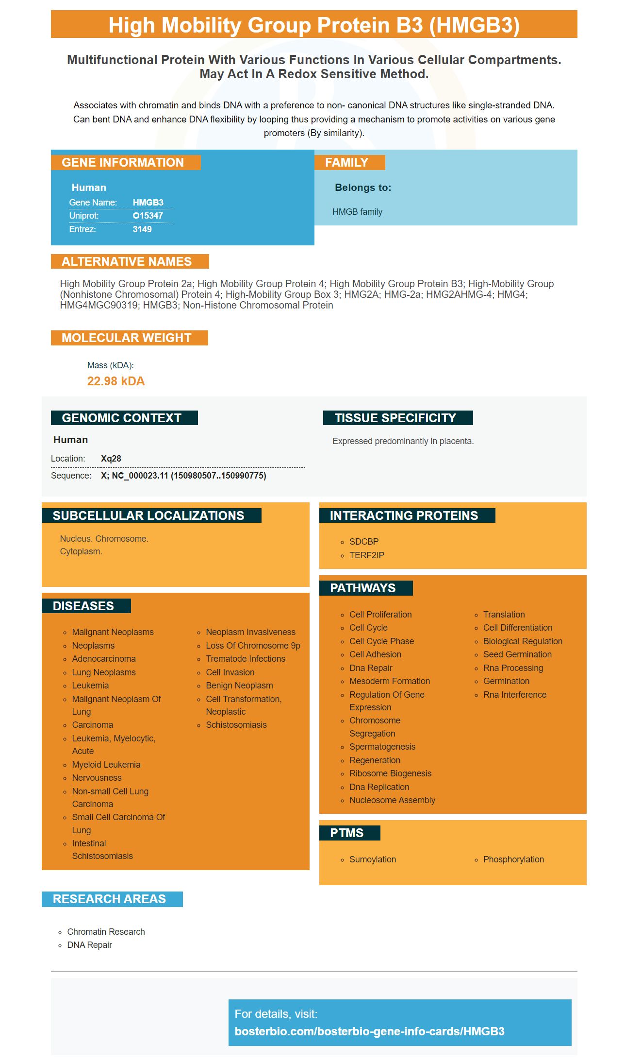
Facts about High mobility group protein B3.
Associates with chromatin and binds DNA with a preference to non- canonical DNA structures like single-stranded DNA. Can bent DNA and enhance DNA flexibility by looping thus providing a mechanism to promote activities on various gene promoters (By similarity).
Human | |
---|---|
Gene Name: | HMGB3 |
Uniprot: | O15347 |
Entrez: | 3149 |
Belongs to: |
---|
HMGB family |
High mobility group protein 2a; High mobility group protein 4; high mobility group protein B3; high-mobility group (nonhistone chromosomal) protein 4; high-mobility group box 3; HMG2A; HMG-2a; HMG2AHMG-4; HMG4; HMG4MGC90319; HMGB3; non-histone chromosomal protein
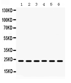
Mass (kDA):
22.98 kDA
Human | |
---|---|
Location: | Xq28 |
Sequence: | X; NC_000023.11 (150980507..150990775) |
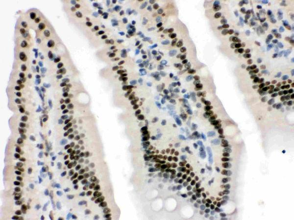
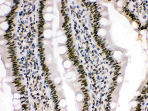
Expressed predominantly in placenta.
Nucleus. Chromosome. Cytoplasm.
The HMGB3 protein is expressed from E. coli and contains a His-Tag and a 1-180 aa sequence domain. This marker can be stored at +2degC to +8degC for a week, or -20degC to -80degC for long-term storage. It is also applicable to all scientists around the world.
HMGB proteins are components of higher order chromatin structures and are involved in binding DNA and cellular RNAs. They are also critical in regulating genome processes in various disease states. However, their exact functions remain unclear. This article will discuss the role of HMGB proteins in the regulation of gene expression. We'll discuss how these proteins regulate DNA methylation, acetylation, and phosphorylation.
Although HMGB4 is found in all tissues, its expression in the somatic cell is restricted. In the embryonic mouse, its mRNA is undetectable at E10.5, but it is highly expressed in the brain, pancreas, and testes by E14. However, in mammals, HMGB4 expression is highest in the testes, where elongated spermatocytes and round spermatids are produced. The expression of the HMGB4 gene is low in other tissues, such as the kidney.
Despite the importance of HMGB4 in the development of the embryo, it is unknown whether it is a therapeutic target. Recent studies suggest that nuclear HMGB1 may serve as a biomarker for cancer. Its level in cancer cells reflects the proliferative state of the cell. Most studies of nuclear HMGB proteins have been conducted in cell and in vitro systems. Future studies may include in vivo models. A conditional knockout mouse has been developed to facilitate these studies. Only time will tell what the HMGB proteins can do.
In addition to being a potential tumor suppressor, HMGB1 affects telomerase activity. This protein co-localizes with telomere binding protein TRF1 and is recruited to telomeres by ChIP. Knockdown of HMGB1 in mice results in increased DNA damage moutelomere ends. Telomerase also contains a DNA template, which complements single stranded DNA overhangs. Further research is needed to determine whether the HMGB proteins play a role in telomere homeostasis.
Unlike HMGB3 and HMGB4, human HMGB2 is structurally defined, whereas HMGB3 and BMGB3 lack an acidic tail. Human HMGB2 contains two homologous DNA-binding motifs, HMG boxes A and B, and an acidic C-terminal domain. Basic amino acids connect these two regions, while acidic amino acids make up the C-terminal region.
Although both HMGB1 and HMGB3 share over 80% similarity in amino acid sequence, HMGB3 lacks an acidic end. However, they are characterized by a long C-terminal acidic tail. They are both transcriptional repressors, but they differ slightly in their composition. This makes mass spectrometry an attractive choice for protein identification. But the problem arises when determining which HMGB protein is which.
The enzyme's ability to inhibit cell proliferation has been investigated in a variety of cancer types. In a recent study, HMGB3 expression was associated with shorter OS in patients with BRCA mutations, whereas HMGB2/3 exhibited a positive correlation with short-term survival. Further research is required to determine the role of HMGB2 and HMGB3 in hepatocellular carcinoma.
Mutations in HMGB1 have been shown to inhibit RAGE activity. Mutants that lack glycines in amino acids 102-105 show an enhanced ability to bind cytokines such as IL-1b. CpG DNA also enhances HMGB1's ability to bind RAGE. These mutations are necessary for RAGE activation, and a HMGB3 lacks an acidic tail prevents this from occurring.
HMGB1 is the only nuclear HMG protein known to activate RAGE. No other nuclear HMG proteins have been shown to activate RAGE. HMGB proteins can complex highly bent, platinated DNA and CpG DNA. Because HMGB1 is abundant in most tissues, it's available for translocation to the nucleus. In addition, HMGB2 and HMGB3 are upregulated in some cancers, suggesting that they may play a role in activating RAGE.
HMGB3 is a transcription factor that controls the expression of several neuronal differentiation markers. HMGB1 and HMGB2 mRNA expressions were high in proliferating NSCs and decreased at E14.5 and P0. During differentiation, HMGB3 protein expression was unchanged. In contrast, HMGB2 protein expression decreased by more than 10% in differentiating spheres.
HMGB1 and HMGB2 are members of the HMG superfamily. Both proteins bind to nucleosomes and the minor groove of DNA. HMGB1 and HMGB2 are expressed at high levels during the proliferative phase of embryonic telencephalon development and decrease substantially in differentiating NSCs. These findings suggest a role for HMGB1 and HMGB2 in neurogenesis and radial glia development.
HMGB3 is highly active in the EC. It also interacts with c-myc, b-catenin, and MMP7. These pathways contribute to the progress of the embryonic stem cell (EC) stage. HMGB3 is associated with the expression of neuronal differentiation markers such as neurite outgrowth, morphogenesis, and axonogenesis.
Down-regulation of HMGB3 inhibits cell migration and invasion. It also suppresses the expression of the Wnt/b-catenin pathway, MMP7, and c-myc. Western blots and immunohistochemistry analyses revealed that the down-regulation of HMGB3 suppresses cell migration and invasion. It also inhibits the expression of neuronal differentiation markers, such as c-myc.
In addition, HMGB3 represses the Xbra2 promoter, while preserving the Xnr1 and Mix2 promoters. These results indicate that HMGB3 may control a subset of mesoderm genes during the gastrula stage. If these genes are over-expressed, this protein can lead to developmental abnormalities. It is not known how HMGB3 regulates neuronal differentiation markers, but it may have a role in regulating neuronal differentiation.
The HMGB family of proteins is largely conserved between animal species. Plants have higher levels of these proteins in their genome than mammalian counterparts, but their structure differs. Plant HMGB proteins have only one HMG box and one C-terminal flanking acidic region. The genome of A. thaliana codes for 8 HMGB proteins. The N-terminal basic domain enhances DNA binding.
The amino acid sequence of HMGB3 determines its nucleo-cytoplasmic distribution. HMGB is a protein that shuttles between the nucleus and cytoplasm. It co-localizes with histones in the cell nucleus to facilitate gene expression under salinity stress. HMGB3 is not found in chloroplasts or mitochondria.
HMGB3 co-localizes in the cell nucleus. HMGB1 and HMGB3 form a complex that interacts with histones and chromosomes. In Drosophila and mammals, the HMG proteins are associated with the SWI/SNF complexes, the BAF complex, and the histone chaperone FACT.
HMGB proteins function as utility players in the cell nucleus. They play an essential role in nucleosome remodeling. In addition to their role in DNA binding, they also inhibit RNA processing and transcription. In vivo, HMGB1 preferentially binds to damaged DNA, inhibiting RNA and DNA repair by blocking access to the NER proteins.
HMGB3 has been linked to the development of chromosomes. HMG proteins are responsible for the development of many different cell types, including humans. However, HMGB3 also interferes with histone h2 binding. It also prevents the formation of new chromatin. In the nucleus, HMGB3 interacts with histones in the cell nucleus.
HMGB3 has a critical role in the development of cervical cancer. HMGB3 binds to the promoter region of hTERT and activates its transcription. Thus, targeting HMGB3 could provide a useful strategy for cervical cancer treatment. Currently, no drugs targeting HMGB3 have been approved. However, future studies are warranted to test HMGB3's role in cervical cancer.
Moreover, HMGB3 has a profound effect on the replication initiation landscape. This process has prominent SNS enrichments in a cell population. Recently, short strand sequencing has been applied to identify ORIs in several systems. Hence, identifying ORIs in cells expressing mutant HMGB3 may help in the future. These findings provide a more detailed understanding of how HMGB3 regulates chromatin.
The expression of HMGB3 in normal and GC tissues was studied using an online database. In addition, we also examined the relationship between HMGB3 and Meis2/miR-18. The results are shown in the figure. GraphPad was used to analyze the data. The data were expressed as mean + SD (SD), n = n. P values were calculated by t-test.
Despite these promising results, these data do not support the clinical significance of HMGB3 in NB. Although HMGB3 is associated with unfavorable outcomes in NB, HMGB3 has a pivotal role in tumor growth, proliferation, and invasion. Therefore, preventing the expression of HMGB3 could represent a novel therapeutic target for patients with this condition. It may also be useful for predicting NB patient survival and outcome.
HMGB1-KO mice die soon after birth. MEFs made of HMGB1-KO mice are viable but contain reduced levels of canonical histones and H2A.X variants. HMGB3 also regulates nucleosome assembly. Because of the decreased nucleosome number, the nucleosomes are not evenly distributed throughout the genome. Consequently, nucleosomes spend less time in each position. These alterations in chromatin dynamics are associated with increased gene abundance and a specific alterations in the expression of subsets of genes.
PMID: 9370291 by Wilke K., et al. Isolation of human and mouse HMG2a cDNAs: evidence for an HMG2a- specific 3' untranslated region.
PMID: 20598277 by Sun Y., et al. Terminal osseous dysplasia is caused by a single recurrent mutation in the FLNA gene.