This website uses cookies to ensure you get the best experience on our website.
- Table of Contents
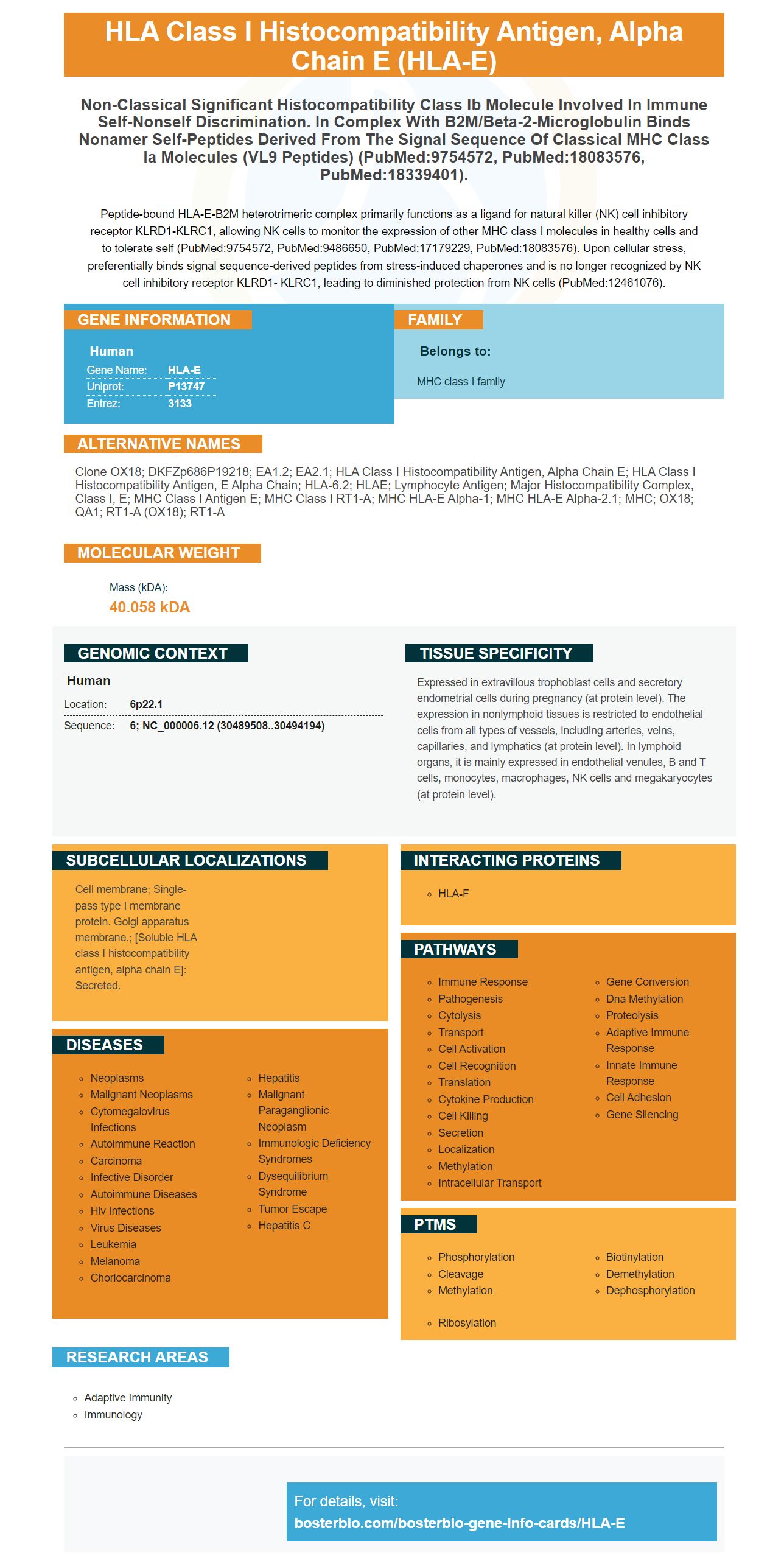
Facts about HLA class I histocompatibility antigen, alpha chain E.

Peptide-bound HLA-E-B2M heterotrimeric complex primarily functions as a ligand for natural killer (NK) cell inhibitory receptor KLRD1-KLRC1, allowing NK cells to monitor the expression of other MHC class I molecules in healthy cells and to tolerate self (PubMed:9754572, PubMed:9486650, PubMed:17179229, PubMed:18083576). Upon cellular stress, preferentially binds signal sequence-derived peptides from stress-induced chaperones and is no longer recognized by NK cell inhibitory receptor KLRD1- KLRC1, leading to diminished protection from NK cells (PubMed:12461076).
| Human | |
|---|---|
| Gene Name: | HLA-E |
| Uniprot: | P13747 |
| Entrez: | 3133 |

| Belongs to: |
|---|
| MHC class I family |

Clone OX18; DKFZp686P19218; EA1.2; EA2.1; HLA class I histocompatibility antigen, alpha chain E; HLA class I histocompatibility antigen, E alpha chain; HLA-6.2; HLAE; lymphocyte antigen; major histocompatibility complex, class I, E; MHC class I antigen E; MHC Class I RT1-A; MHC HLA-E alpha-1; MHC HLA-E alpha-2.1; MHC; OX18; QA1; RT1-A (OX18); RT1-A
Mass (kDA):
40.058 kDA

| Human | |
|---|---|
| Location: | 6p22.1 |
| Sequence: | 6; NC_000006.12 (30489508..30494194) |
Expressed in extravillous trophoblast cells and secretory endometrial cells during pregnancy (at protein level). The expression in nonlymphoid tissues is restricted to endothelial cells from all types of vessels, including arteries, veins, capillaries, and lymphatics (at protein level). In lymphoid organs, it is mainly expressed in endothelial venules, B and T cells, monocytes, macrophages, NK cells and megakaryocytes (at protein level).
Cell membrane; Single-pass type I membrane protein. Golgi apparatus membrane.; [Soluble HLA class I histocompatibility antigen, alpha chain E]: Secreted.


We will discuss HLA-E mAbs and Mock controls, Immunohistochemical analysis, and the clinical implications. We will also examine how this mAb can be used in cancer cells. We will also discuss potential applications for HLA-E immune therapy. HLA E mAb is available in purified and Recombinant forms.
Boster Bio provides a vector for HLA expression that allows researchers create stable transfected cells BUF1088 cells that are overexpressed with the human antigen. They can then measure the HLA protein level in cells by using intracellular flow and Western blotting. These cells can then be compared to mock transfected control cells to determine if they are functionally transfected.
The gene encoding the nonclassical human leukocyte antigen E is overexpressed in various tumor types, including human renal cell carcinoma. It is an important immunomodulatory molecule and may represent a key pathway to tumor escape. However, it is not known if HLA-E expression is involved in human renal cell carcinoma. HLA-E expression is strongly correlated with tumor size.
HLA E expression increases are associated with tumor progression and metastasis formation. HLA-E expression in some tumors was associated with lower survival rates. Additionally, high levels of HLA-E surface expression were found on hematopoietic malignancies, including multiple myeloma and leukemia. It was also associated with decreased cytotoxicity and a reduced NK cell cytolysis.
Immunohistochemical staining of TMA-M sections using specific mAbs for NK cells, T cell subsets, and activation markers indicated a correlation with HLA-E expression. Additionally, ionizing radiation increased the level of HLA-E in GBM cells. These results suggest that HLA-E could be used as a biomarker to evaluate the effectiveness of treatments.
In addition, HLA-E protein induction was not induced in HEK293T cells by IFN-g. The MEM-E/02 antibody also showed cross-reactivity to classical HLA class Ia compounds. This suggests that this antibody may have high cross-reactivity with other HLA-E molecules. And this is just a sample of Boster Bio's HLA-E expression vector.
In addition to developing ELISA kits, Boster Bio also develops research antibodies. Boster Bio's antibodies are able to detect biomarkers that can be used to diagnose cancer, neurological disorders, inflammation, and development. These antibodies have been validated against a panel of 250 tissues, including untransfected cell lines. They also have excellent affinity. Tebu Bio also has their immunological products. They are supported by Sanbio’s technical support.
The transient transfection of the HLA-E gene with an HLA–E expression vector was used to determine the functional HLA/E expression in HEK293T. To perform the staining we used a specific anti-HLA-E antibody (TFL033) to detect intracellular HLA–E.
A mAb called 3H4 specifically binds the a2 domains of HLA–E and recognizes them. It did not bind either to the mouse orthologs nor to the rhesus. These results suggest that 3H4 recognizes HLA E VL9 complex. Lastly, threeH4 recognizes the human and mouse HLA-E-VL9 tetramer.
We created pan-B cell lines from human pans by negative selection. Then, we used a three-color sorting strategy to distinguish the HLA-E-VL9 marker from the control cells. HLA E-VL9 doublepositive B cells showed that pan-B cell sorting was more efficient when they were HLA E-VL9 positive. Next, we isolated variable regions from the sorted cell using PCR. These regions were then cloned together with the human IgG1 constant to create an expression backbone.
Then, we generated a PCR amplification of the HLA-E heavy chain using the pcDNA3.1 vector (Invitrogen). The forward primer contained the last repeats from the (G4S.4)4 linker. The LGC genomics protocol was used for the synthesizing of the fragment. The gB2M targeting site was changed to 5'-CCGTGTGTGTGTGTGTACTC-3'.
The binding of the HLAE heavy chain to peptide is accomplished by the VL CDR3 C-junction. The HLAE peptide-binding groove is formed by surface loop residues that sweep across the groove. The HLA-E a1 helix R62 is colored marine blue, while the residues involved in the 3H4 CDR3 D-junction are color coded.
The Mock control of the HLA-E gene provides the opportunity to determine the functional role of tapasin. Tapasin is responsible for the assembly of class 1 molecules in the ER. The HLA-E gene is transiently expressed at the cell's surface. HLAE, which encodes HLAE, is highly conserved in human genome.
The HLA-E protein is expressed on many different types of tumor cells and is a powerful marker of a number of neoplastic diseases, including CRC. These cells can be detected using immunohistochemical staining. The procedure involves the use of a mouse monoclonal antibodies to HLA-E. The primary antibody is incubated in the cells for 45 min at 4degC. 30 minutes later, the secondary antibody is added. The average intensity level refers to the percentage of cells that stain positive for a particular colored color.
HLAE protein is present in low levels in normal epithelial cell tissue, but it is abundant in the stroma. To determine the extent of stroma, the tumor cells were compared against a control group of infiltrating immune lymphocytes. A dot plot was used in order to determine the percentage HLA-E positive cell.
High levels of immunocytochemical staining was used to identify tumor cells that had high levels HLA E protein. The likelihood of lymphocytes being present in tumors with high levels HLA-E expression was significantly lower than normal tissue. Internally, lymphocytes infiltrating from the skin were used as a positive control. The percentage of HLAE positive cells in tumours in the Dukes' B and C grades was 72.4+-3.0 compared with 6.2+-1.2 in normal mucosae.
Prof. Dr. J. Neefjes was the one who derived the antibodies used to detect HLA E. They recognize HLA-E denatured and peptide-free monomers. The TFL033 antibody was used as negative control in the study. The intracellular HLA E a chains were stained by the TFL-033 antibody.
HLA-E is an MHC class I nonclassical molecule that has recently been identified as a potential marker for advanced tumor stages and disease progression. Recent studies have linked HLA-E expression to poor outcomes in solid tumors. However, there is still no clear evidence as to whether HLA-E expression can impact the outcome of a transplant. We studied 110 patients with chronic lymphocyticleukemia to test this hypothesis.
Although HLA E's functions are not yet known, this molecule is vital for both adaptive and innate immune reactions. Because of its low polymorphism and relative insensitivity to downregulation by HIV, HLA-E can be used as a target in vaccination strategies. HLA-E-restricted HLA-E T-cells should be able to mount effector responses and recognize foreign antigens. Further research is needed to determine the role of HLA/E in these functions.
RCC contains the HLA-E marker, which is associated with decreased immunogenicity. Although the study's findings have important clinical implications it is still insufficient information to draw conclusions. Future studies are needed to better understand the role played by the HLA-E marker in different types of cancer. Its use in diagnosis of cancer is just one potential application. This molecule does not have a reduced chance of survival from specific diseases.
The expression of HLA-E was not significantly correlated with the immune cell markers CD3 or CD8. However, it did show an inverse correlation to the infiltration NK-cells. In fact HLA-E was inversely correlated to the number NK cells infiltrating RCC. However, the correlation was not significant with other cytokine markers like CD4, FOXP3,CD69, or CD25.
Although there is not yet any evidence that HLA-E has an effect on the immune response, it has been suggested HLA-E modifications may be beneficial in certain situations. In particular, HLA-E-dimorphism decreases NK cell activity and reduces IL-2-mediated cytotoxicity. Further studies are needed to confirm the effects of this mutation on NK cells and to determine whether it can be used in other conditions.
HLAE-expression has been found in many tumors and cell lines. However, it has not previously been studied to determine if HLAE-expression is associated with overall survival in RCC patients. The markers are associated with chromophobe RCC and NK cells, but are not significantly correlated with WHO tumor grading or immunotherapy response. HLA–E plays an important role in RCC. Although it may play an important role for the immune surveillance of tumors and other aspects of cancer, further research is needed to determine if HLA E-overexpression is a contributing factor.
PMID: 3131426 by Mizuno S., et al. Isolation and nucleotide sequence of a cDNA clone encoding a novel HLA class I gene.
PMID: 10064069 by Ulbrecht M., et al. Cell surface expression of HLA-E: interaction with human beta-2 microglobulin and allelic differences.

