This website uses cookies to ensure you get the best experience on our website.
- Table of Contents
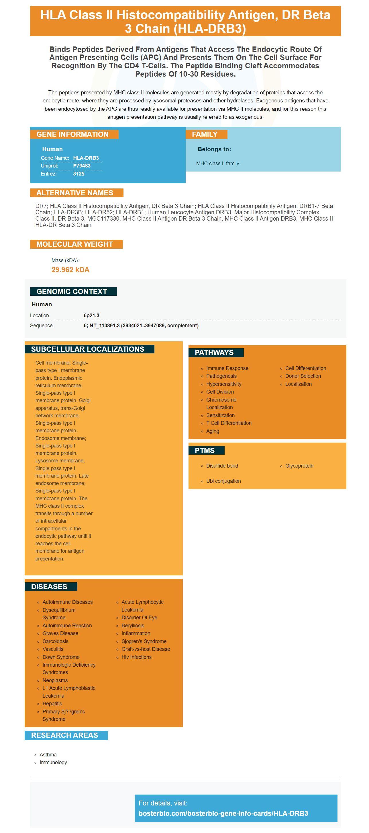
Facts about HLA class II histocompatibility antigen, DR beta 3 chain.
The peptides presented by MHC class II molecules are generated mostly by degradation of proteins that access the endocytic route, where they are processed by lysosomal proteases and other hydrolases. Exogenous antigens that have been endocytosed by the APC are thus readily available for presentation via MHC II molecules, and for this reason this antigen presentation pathway is usually referred to as exogenous.
Human | |
---|---|
Gene Name: | HLA-DRB3 |
Uniprot: | P79483 |
Entrez: | 3125 |
Belongs to: |
---|
MHC class II family |
DR7; HLA class II histocompatibility antigen, DR beta 3 chain; HLA class II histocompatibility antigen, DRB1-7 beta chain; HLA-DR3B; HLA-DR52; HLA-DRB1; human leucocyte antigen DRB3; major histocompatibility complex, class II, DR beta 3; MGC117330; MHC class II antigen DR beta 3 chain; MHC class II antigen DRB3; MHC class II HLA-DR beta 3 chain
Mass (kDA):
29.962 kDA
Human | |
---|---|
Location: | 6p21.3 |
Sequence: | 6; NT_113891.3 (3934021..3947089, complement) |
Cell membrane; Single-pass type I membrane protein. Endoplasmic reticulum membrane; Single-pass type I membrane protein. Golgi apparatus, trans-Golgi network membrane; Single-pass type I membrane protein. Endosome membrane; Single-pass type I membrane protein. Lysosome membrane; Single-pass type I membrane protein. Late endosome membrane; Single-pass type I membrane protein. The MHC class II complex transits through a number of intracellular compartments in the endocytic pathway until it reaches the cell membrane for antigen presentation.
There are many options to choose from when setting up a flow method. These tips and guidelines can help optimize your experiment no matter what choice you make. Learn more in the following article. It contains information on the NS5551-565 peptide (epitope), and tetramer. These tools should be used correctly to achieve the best results. This marker has many uses.
The NS5551-565 epitope is a part of the NS5552-565 tetrapeptide molecule, which is highly expressed in tumor cells. Boster Bio develops high-sensitivity, high-specific antibody kits for many research applications, including immunology, neuroscience, and cancer. Boster Bio's antibodies are tested for IHC, WB, Flow Cytometry and other diagnostics.
This epitope is recognized by T cells through the HLA-DRB1*03:01 and HLA-DRB3*03:01 restriction elements. However, this epitope's non-protruding ends are not altered in EnvE4458 donors. This result suggests that additional restriction factors may be present within the human tumor cell lineage.
The CD8+ T-cell epitope NS5551-565 has a predicted binding affinity between 0.8 and 1.3. Initial identification of the epitope was HLA-A-restricted and HC-B. Its length was determined using reverse immunology methods and HFRI. These produced 10 unique HLA–B-restricted, CD8+ T-cell epitopes. The same method was used to produce eight additional CD8-T cells tetramers using the same approach.
Boster Bio researchers discovered the NS5551-565 epitope in 2010. The peptide had been presented to the T-cell lineage that was restricted by multiple HLA/DR types. Although no donors had three HLA DR molecule molecules, some donors could generate CD4+ cells responses that were restricted only by one of the restriction elements. The researchers used a biochemical binding affinity test to determine if both tetramers had the same epitope.
HLA-DRB1*15.01, HC-DRB5*01.01. also present the NS5551-565 Epitope. The YF1067 library of peptides contains 31 CD4 + T cells epitopes, which can be validated with tetramers. This method can identify NS5551-565 cancer immunotherapy epitopes by reducing the number and complexity of combinations.
There are 13,610 peptides total from the YFV protome. Both NetMHCpan 4.0EL and MixMHCpred both have a high rate false-positive predictions. This is a sign of the effectiveness of these antigen prediction tools in high-sensitivity and high affinity immunoassays. The YFV NS4B 214-222 epitope, which is an immunodominant antigen, is a good example. All 93 HLA+A-positive donors responded positively to the epitope.
HLA-DRB3 presents the NS5551-565 peptide as a tetramer, with the marker HLA–DRB3 * 13. The tetramers contained the overlapping 'peptides in a 15-mer couple and were stained to confirm their specificity.
To verify that the peptide could be effective against CD8+ t cells, the tetramers of the peptide were tested against the HLA DRB3*01/15:01 molecule. The peptides corresponding each intersection were identified by a four-matrix ICS. 27 of the resulting HLA combinations were confirmed to be CD8+-T cell stimulatory and peptides.
The NS5551-565 peptide targeted both CD4+ andCD8+ T cells. It was capable of recognizing both CD4+ and CD8+ T cell in one test. This process may allow the development of a specific drug to combat a given disease. The HLA-DRB3 and NS5551-565 peptides are not yet approved for clinical trials.
Researchers need to identify the peptides binding to the human MHC–DRB3 protein in order to create a high-quality antibody. Sette et al. analyzed a group of single HLA-II-transfected lines of cells. A tetramer of 16 HLAB*07:02+ donors was created.
The antibody targets HLA and DRB3 restricted elements. It was highly specific in testing both types. The HFRI approach was more sensitive than the HFRI screen strategy. However, it was more specific and sensitive. It also had a higher rate of sensitivity. It was also less specific. It can be used as a marker for HLA DRB3-restricted patients.
The overlapping peptides that represent the yellow fever virus proteome were screened for peptides containing CD4+ T cell epitopes. The peptide-HLA tetramer can be used to validate T cell epitopes, which reduces the false discovery rate. The HLA DRB3 marker can also be found in the overlapping proteins, which allows the Tetramer to be used in a wider variety of applications.
Boster Bio's NS5551-565 tetrapolymer recognizes a wide variety epitopes. Boster's antibodies have been extensively referenced in peer-reviewed journals for the past 25 years, which demonstrates their value to the research community. Several of their antibodies have been validated in ELISA, Western Blotting, and Immunohistochemistry.
It is also the first tetramer capable of recognizing NS5551-565 (an epitope found on HLA DRB1*01, HLA DRB5). The tetramer has different flanking regions, but it binds to core epitopes on HLA-1*01 and HLA-5. These results suggest that diversification is essential for protecting the body from pathogens. Another validation is needed in order to determine how many CD4+T cell epitopes exist on human immune systems.
PMID: 11894954 by Long E.O., et al. Complete sequence of an HLA-DR beta chain deduced from a cDNA clone and identification of multiple non-allelic DR beta chain genes.
PMID: 3459965 by Gorski J., et al. Polymorphism of human Ia antigens: gene conversion between two DR beta loci results in a new HLA-D/DR specificity.