This website uses cookies to ensure you get the best experience on our website.
- Table of Contents
1 Citations 1 Q&As
1 Citations
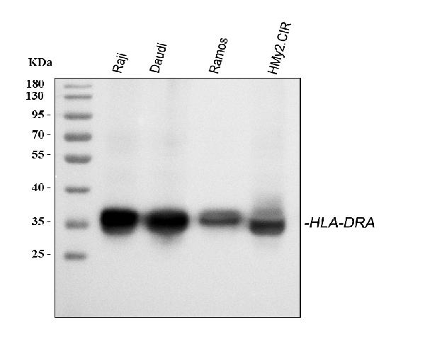
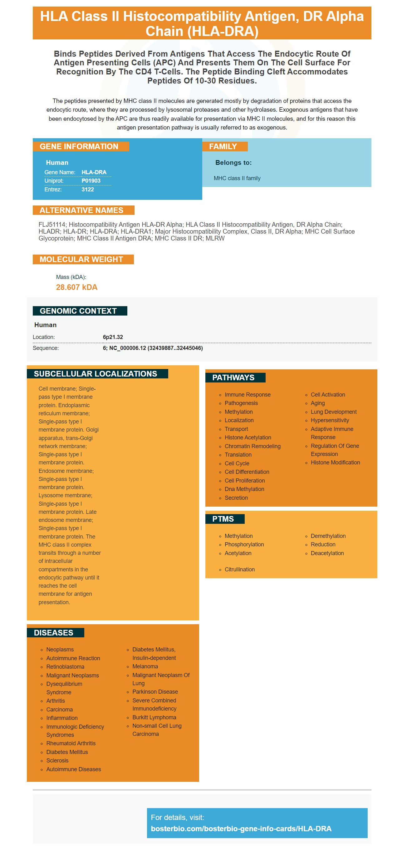
Facts about HLA class II histocompatibility antigen, DR alpha chain.
The peptides presented by MHC class II molecules are generated mostly by degradation of proteins that access the endocytic route, where they are processed by lysosomal proteases and other hydrolases. Exogenous antigens that have been endocytosed by the APC are thus readily available for presentation via MHC II molecules, and for this reason this antigen presentation pathway is usually referred to as exogenous.
Human | |
---|---|
Gene Name: | HLA-DRA |
Uniprot: | P01903 |
Entrez: | 3122 |
Belongs to: |
---|
MHC class II family |
FLJ51114; histocompatibility antigen HLA-DR alpha; HLA class II histocompatibility antigen, DR alpha chain; HLADR; HLA-DR; HLA-DRA; HLA-DRA1; major histocompatibility complex, class II, DR alpha; MHC cell surface glycoprotein; MHC class II antigen DRA; MHC Class II DR; MLRW
Mass (kDA):
28.607 kDA
Human | |
---|---|
Location: | 6p21.32 |
Sequence: | 6; NC_000006.12 (32439887..32445046) |
Cell membrane; Single-pass type I membrane protein. Endoplasmic reticulum membrane; Single-pass type I membrane protein. Golgi apparatus, trans-Golgi network membrane; Single-pass type I membrane protein. Endosome membrane; Single-pass type I membrane protein. Lysosome membrane; Single-pass type I membrane protein. Late endosome membrane; Single-pass type I membrane protein. The MHC class II complex transits through a number of intracellular compartments in the endocytic pathway until it reaches the cell membrane for antigen presentation.
If you've been studying the HLA-DRA marker, then you have probably heard a lot of hype. This molecule, an alphabeta-chain heterodimer is linked to the hepatitis B viral clearance. Its purpose is to help the immune system recognize and respond to foreign substances and antigens. These are the main applications of this marker in research.
The function of the HLA DR molecule is not clear. It inhibits the presentation of antigens in MHC class II compartments. It is also expressed primarily in B cells. It could be a repressor to class II peptides, or a cell-specific modulator. It could also play some role in the maintenance of the repertoire of antigens that are released by B cells.
The endoplasmic retina is home to MHC II complexes. The invariant chain guards them from binding to cellular proteins. These complexes then escape through the Golgi apparatus which exchanges the peptides that have been attached to them for foreign antigens. HLA-DM chaperones a majority of APC molecules and HLA-DO chaperones B cells.
The unique arrangement of three genes in the human HLA system codes for the three main types of MHC molecules. The a-chain gene is organized in a noncovalent manner and the b-chain gene is composed of one exon that encodes the transmembranedomain and another that codes for the cytoplasmic tail.
HLA-DR is a molecule which can be utilized for multiple purposes. The protein is responsible for the control of immune responses . It also functions as a marker of activation of T cells. This molecule is frequently associated with disease activity. It is also associated with HIV infection. It is important to remember that the HLA-DR is one of the genes that are associated with autoimmune diseases and rheumatoid disease.
HLA-DR expression was observed in T and B cells in this study. Patients with HLA-DR negative disease did not detect MHC-DR-specific expression on monocytes or lymphocytes. However the molecule was detected on specific cells which included professional and innate lymphoid cell types.
The paralogue of HLA B, HLA-DRA, is second most important human gene. It is responsible to present extracellular proteins in the form peptides. The alpha chain spans 33 to 35 kDa in size and includes five exons. The DRA alpha chain does not contain polymorphisms inside the peptide-binding region and functions as the sole alpha chain of DRB1, DRB3, and DRB4.
The LD is most intense in small blocks. The region that contains HLA/DR/DQ loci has 1.2 Mb of long-range LD (Fig. 2). In the same region in the same region, associations with the HLA DO gene depended on the presence of the HLA-DQB1 SNV. The PRRT1-related associations were also conditional on the presence of the HLA-DO gene and a TAP1 gene located in the vicinity of HLA-DQB1.
The a3-domain of the HLA E gene is responsible for the creation of the gene. HLA-E is closely related to HLA A and HLA B due to eight sites in the following 99 nucleotides. However HLA-E is more closely associated with HLA-B and HLA -C than it is to HLA-D.
The HLA-DRA gene encodes for a heterodimer of HLA class II alpha chains. The alpha chain binds a specific peptide, which could be anywhere from ten to thirty residues in length. The peptides generated by lysosomal enzymes and other hydrolases are readily accessible to MHC II molecules. The immune system and immune response are influenced by the HLA-DRA molecule.
Although these molecules are identical, they aren't identical. They have some commonalities. HLA-DRA is found in many tissues, including the spleen, the skin and lungs, as well as the skin. Researchers are currently studying the relationship between HLA-DRA and other immune system proteins. This is the basis for developing immunogens for research purposes. These antibodies are made in a lab that is specially designed to ensure the highest quality.
It has been widely believed that the alleles HLA-DRB1*11/12 and HLA-DRB1*07 are associated with hepatitis B virus (HBV) persistence and infection. A study concluded that the DRB1*11 allele was associated with a decreased risk of chronic HBV infection , whereas HLA-DRB1*12 was not. Although the results from the study aren't conclusive, the findings support that these alleles are associated with a lower risk of HBV infection and persistence.
Participants in the study were at least 40 years old age and were seronegative against anti-HBV, HBV and anti-HCV serological markers. The controls, or hypernormals were negative for all tests, however they had at least two consecutive positive HBV tests results without any evidence of cirrhosis. These patients were described as "hypernormal" by researchers to differentiate them from the general population.
The study also showed that certain genotypes of HLA-DRA are associated with the hepatitis B virus infection. These studies have demonstrated that different variants of HLADRA have different effects on progression of the disease. These findings will aid in the development of specific, personalized treatment and prevention strategies for chronic HBV infections. Genetic testing is now a standard part in the treatment of HBV infection.
There are currently around 400 million people in the world with HBV infection. Around 10 percent of chronic HBV infections lead to Hepatocellular Carcinoma (HCC) while a smaller number of chronic HBV infections are affected. It is important to know that the susceptibility of a person to HBV infection is multifactorial as host immunization status, genetic factors and HBV subtypes playing an integral role.
Researchers discovered three motifs in the HLA-DRB1 gene of humans that could be associated with T1D. The motifs are known as "KAG", "EAV" and RAG. The other three motifs are related to reduced risk of T1D. The HLA-DRB1 protein is able to bind peptides with KAG motif but not. This motif has been associated with T1D, APS3v susceptible, and APS3v susceptible regions.
The HLA-DRA molecule can also be an option for diagnosing of T1D and other autoimmune disorders. It targets autoantigens of various kinds and has a distinct pocket amino acids signature. This particular motif triggers specific structural modifications in the MHC II peptide binding pocket that affect the peptide binding and the presentation of antigens. The development of many immune-mediated diseases is dependent on the amino acids arg-74. Blocking this pocket may offer a therapeutic approach for certain T1D patients.
There is no clinical test to test for T1D using the HLA-DRA gene. However, recent studies have revealed that non-classical HLA genes play a role in the development of T1D. Morgan et al. concluded that the detection of the HLADRA gene in blood samples could help doctors identify the condition and help improve treatment.
The gene is located in 148 proteins-coding genes. There are a variety of genes that contribute to immune responses and cell processes. Using a statistical model, the HLADRA gene was associated with the development of T1D with NDR-defined T1D. The genome is comprised of more than the 20,000 genes, and it's estimated that approximately five hundred thousand of them reach genome-wide significance.
PMID: 6811954 by Lee J.S., et al. Sequence of an HLA-DR alpha-chain cDNA clone and intron-exon organization of the corresponding gene.
PMID: 6416803 by Kajimura Y., et al. Cloning the heavy chain of human HLA-DR antigen using synthetic oligodeoxyribonucleotides as hybridization probes.
*More publications can be found for each product on its corresponding product page