This website uses cookies to ensure you get the best experience on our website.
- Table of Contents
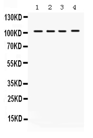
Facts about Histone deacetylase 7.
Involved in muscle maturation by repressing transcription of myocyte enhancer factors like MEF2A, MEF2B and MEF2C. During muscle differentiation, it shuttles to the cytoplasm, allowing the expression of myocyte enhancer factors (By similarity).
Human | |
---|---|
Gene Name: | HDAC7 |
Uniprot: | Q8WUI4 |
Entrez: | 51564 |
Belongs to: |
---|
histone deacetylase family |
DKFZP586J0917; EC 3.5.1.98; HD7; HD7A; HDAC7AFLJ99588; histone deacetylase 7; Histone deacetylase 7ADKFZp586J0917
Mass (kDA):
102.927 kDA
Human | |
---|---|
Location: | 12q13.11 |
Sequence: | 12; NC_000012.12 (47782722..47820612, complement) |
Nucleus. Cytoplasm. In the nucleus, it associates with distinct subnuclear dot-like structures. Shuttles between the nucleus and the cytoplasm. Treatment with EDN1 results in shuttling from the nucleus to the perinuclear region. The export to cytoplasm depends on the interaction with the 14-3-3 protein YWHAE and is due to its phosphorylation.
Interested in learning more about the HDAC7 gene? This article will cover RT-qPCR evaluation of the HDAC7 gene, its application in OA, and how to use it in education. Educators may also share this resource with their students, but they must link to Boster Bio. If you'd like to learn more about the HDAC7 gene, please visit Boster Bio.
RT-qPCR assays have recently been utilized to examine the effects of HDAC inhibitors on cancer stem cells. In the present study, we show that the treatment of cancer stem cells with an HDAC inhibitor increased the activity of OCR. This activity is a hallmark of cancer stem cells and chemoresistance cells. To further explore the role of HDAC inhibitors, we performed a metabolomics analysis.
Using the gene b-2-microglobulin, we found that HDAC7 mRNA expression correlated to the presence of two reference loci. The stability of b-2-microglobulin (MHI), a component of major histocompatibility complex I (MHC), was not significantly different among the three reference loci. Moreover, the expression level of ribosomal protein L13A, an important gene for transcription, was not significantly different among the two reference loci. Therefore, RT-qPCR evaluation of HDAC7 gene expression is highly relevant to the characterization of cell culture tumors.
Using high-throughput RT-qPCR assays based on the Fluidigm BioMark system allows for up to 9216 qPCR reactions at once. These techniques allow for more refined analysis and measure modulation of innate immune gene networks. The results are highly encouraging. This study supports our previous observations regarding the function of HDAC inhibitors in GBM cells. We hope to see further progress in this field.
This study was conducted using PerkinElmer COVID-19 Test Home Collection Kit. Afterwards, we used PerkinElmer COVID-19 RT-qPCR Reagent Kit for the detection of HDAC7. The two antibodies used were CST 2106S and CPT2 antibody. The results obtained from these assays are consistent with the published results. A second study is planned to test the effects of COVID-19 RT-qPCR.
RT-qPCR analysis of HDAC7 is highly sensitive. RNA interference with pan and selective HDAC inhibitors results in increased c-Myc protein levels and reduced cellular viability. These results suggest that HDAC inhibitors inhibit the production of these proteins. The authors are investigating whether they affect the proliferation of GBM cells by reducing the levels of c-Myc. The results also show that HDAC inhibitors have anti-tumor effects.
RT-qPCR evaluation of HDAC4, a key mediator in the regulation of VEGF expression, was performed using Boster Bio's iTRAQ clone. In this study, HDAC4 was reduced by 47% and Runx2 and VEGF were increased by 3.4 and 3.1 fold, respectively. These results are consistent with other studies, suggesting that HDAC4 inhibits VEGF expression.
The RT-qPCR method evaluated samples for the presence of SARS-CoV-2 and HDAC4. The results indicated 349 true positives and 94 false negatives. The No Template Control was used as a no template control in the PCR and as an extraction negative control. If the oligo mix yields negative results, the RT-qPCR method may have been contaminated. If the oligo mix contains DNA fragments, repeat the analysis with a different sample, paying close attention to the "Warnings and Precautions" section.
RT-qPCR analysis of HDAC4 in Boster Bio cells demonstrated that this gene suppresses EC radioresistance through regulation of miR-146a. Both miR-146a and IRAK1 are targets of HDAC4.
For RT-qPCR analysis of HDAC4 in Boster Bio cells, we performed a miRNA transfection using CS cells. The cells were cultured in EpiLife medium containing 60 uM Ca2+. We then transduced the cells with Lipofectamine 2000. To monitor reporter gene expression, cells were lysed 48 h after transfection.
Using a highly sensitive and specific method, we measured the expression of HDAC4 in human ESCC cells with a high-sensitivity RT-qPCR assay. The resultant data indicated that HDAC4 expression was elevated in radioresistant EC cells and tissues. These findings suggest that HDAC4 may play a role in the radioresistance of esophageal cancer.
Scriptaid increased binding of HDAC5 to MEF2C. This demonstrates that Scriptaid inhibits MEF2C transcription and is also essential for Scriptaid-mediated gene regulation. Scriptaid inhibited the expression of Sost and MEF2C. However, it did not affect basal gene expression. These results suggest that Scriptaid is a potent bone regulator.
RT-qPCR is a method used for the detection of viral RNA in a blood sample. It targets the genome of SARS-CoV-2 virus. It is widely used, but it has significant limitations in reliability and throughput. In addition, its widely-misunderstood terminology has led to much misinformation, including the use of it for diagnostic purposes.
A minus reverse transcription control is included in all RT-qPCR reactions, which checks for contaminating DNA. Normally, primers that flank introns cannot anneal to the template. Thus, if PCR amplification is observed without amplification, then it is likely that the template contains contaminating DNA. This DNA will be used for subsequent analysis.
Single-tube RT-qPCR assays are often used to detect SARS-CoV-2. However, their sensitivity is low because the viral RNA has extensive secondary structure that can affect the efficiency of reverse transcription. For this reason, it is essential to optimize the reaction conditions to avoid false-negative results. For example, variable sequences in the ORF1ab gene may lead to low sensitivity.
RT-qPCR is a powerful tool to assess the levels of HDAC8 in a patient's blood. By analyzing the HDAC8 levels in a sample, you can identify the HDAC8 protein in cells and in blood samples. This can be done with just one sample or multiple samples. The results from multiple experiments are then shared and interpreted. This tool has been approved by the FDA.
RT-qPCR is a popular method for evaluating the level of HDAC8. It is used in many applications, including gene expression analysis, RNAi validation, microarray evaluation, and disease research. By analyzing HDAC8 levels in cells, you can identify the specific gene responsible for the condition of your patient. The Boster Bio RT-qPCR evaluation of HDAC8 in patients with chronic diseases.
RT-qPCR is a versatile diagnostic tool, and it can be used on thousands of samples per day. The Boster Bio HDAC8 kit allows you to run the assays simultaneously, reducing the time required for each sample. The software provides you with a downloadable software tool and a detailed manual that explains the process step-by-step. You can then compare the results using a cross-validated reference for accuracy.
Recent advances in platelet-rich plasma (PRP) applications in OA have highlighted the potential for regenerative injection therapy. The resulting regenerative injections may enhance cartilage cell regeneration and may be used in the treatment of OA. However, the application of platelet-rich plasma in OA clinical trials must be carefully monitored, as some of its growth factors have detrimental effects on OA joints. Further, future directions in PRP application in OA may involve searching for a safe and innocuous anti-VEGF antibody.
EVs-miRNAs have potential therapeutic applications in OA, but the current research on the topic may not lead to clinical transformation. Further research is needed to determine the factors that influence the efficacy and safety of EVs in OA. In addition, EVs-miRNAs may be capable of delivering other molecules, such as cytokines. Regardless of the limitations, the clinical transformation of EV-miRNAs enriched in EVs may be speeded up.
Among the 38 compounds that showed promising results, six of them were capable of acting on more than one target. These compounds included eugeniin, 6-hydroxykaempferol-7-O-glucoside, folic acid, neocarthamin, and safflor yellow A. Using these compounds, we were able to identify a number of ligands with broad-spectrum inhibitory activity towards a variety of OA target enzymes.
The applicants filed OA No. 180/927/2014. The Hon'ble Tribunal disposed of the OA within three months. Both Shri Neeraj Agrawal and Shri K.P. Singh appeared for the applicants. Interestingly, the application was disposed of by the Jabalpur Bench of the Tribunal within the stipulated time period. Moreover, the tribunal has ordered that the applicant's OA be disposed of within a maximum period of three months.
PMID: 11262386 by Lee H.-J., et al. Tip60 and HDAC7 interact with the endothelin receptor a and may be involved in downstream signaling.
PMID: 11466315 by Fischle W., et al. Human HDAC7 histone deacetylase activity is associated with HDAC3 in vivo.