This website uses cookies to ensure you get the best experience on our website.
- Table of Contents
2 Citations 17 Q&As
2 Citations 16 Q&As
1 Citations 16 Q&As
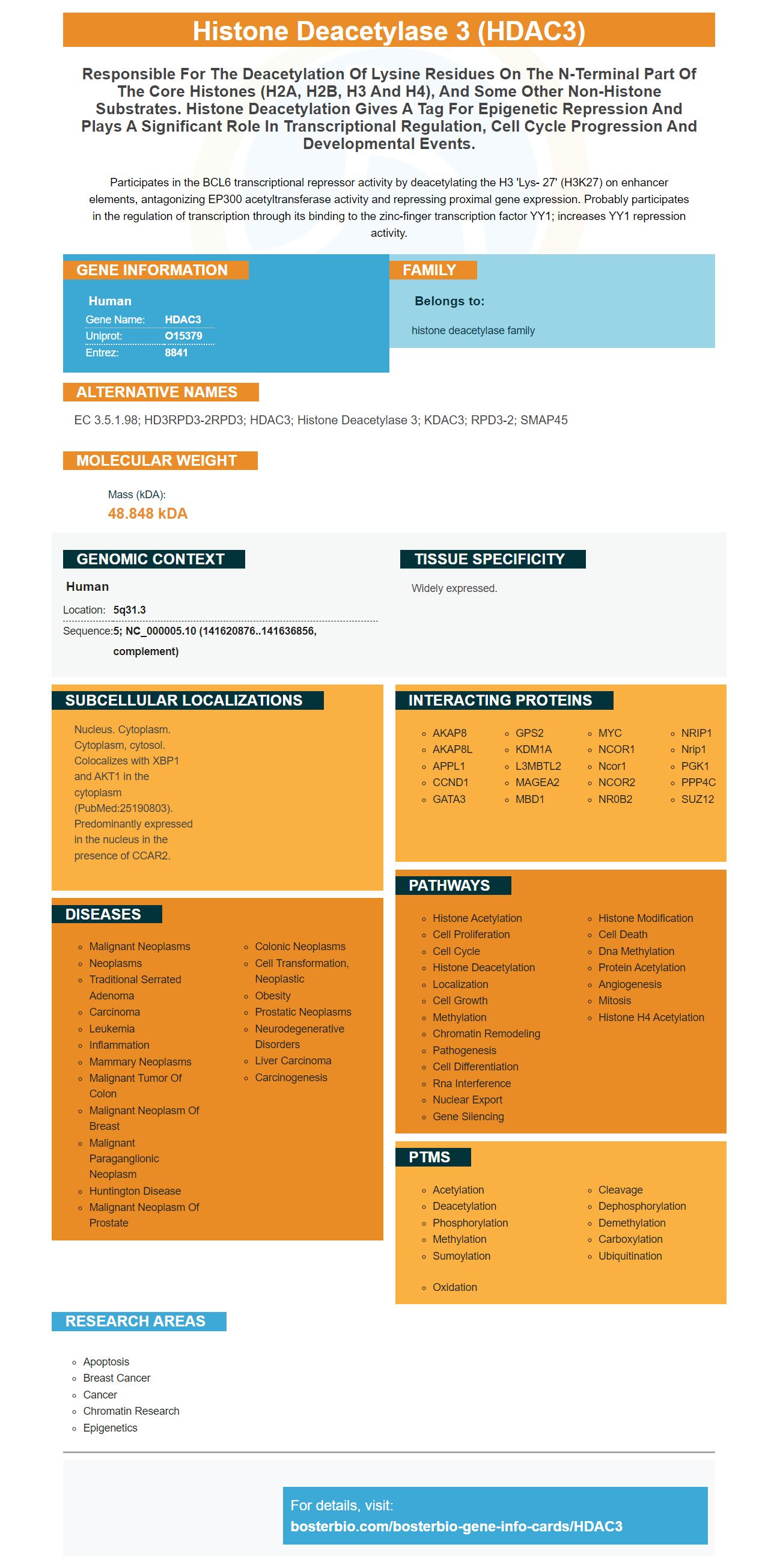
Facts about Histone deacetylase 3.
Participates in the BCL6 transcriptional repressor activity by deacetylating the H3 'Lys- 27' (H3K27) on enhancer elements, antagonizing EP300 acetyltransferase activity and repressing proximal gene expression. Probably participates in the regulation of transcription through its binding to the zinc-finger transcription factor YY1; increases YY1 repression activity.
Human | |
---|---|
Gene Name: | HDAC3 |
Uniprot: | O15379 |
Entrez: | 8841 |
Belongs to: |
---|
histone deacetylase family |
EC 3.5.1.98; HD3RPD3-2RPD3; HDAC3; Histone Deacetylase 3; KDAC3; RPD3-2; SMAP45
Mass (kDA):
48.848 kDA
Human | |
---|---|
Location: | 5q31.3 |
Sequence: | 5; NC_000005.10 (141620876..141636856, complement) |
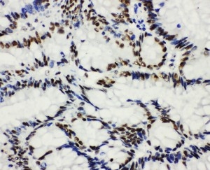
Widely expressed.
Nucleus. Cytoplasm. Cytoplasm, cytosol. Colocalizes with XBP1 and AKT1 in the cytoplasm (PubMed:25190803). Predominantly expressed in the nucleus in the presence of CCAR2.
This article will focus on the HDAC3 marker and the NF-kB DNA binding activity and CD40. Boster also provides product credits to researchers who have submitted their research results. The product credits are applicable to all scientists across the world. If you're a researcher, or a business owner, the HDAC3 marker can assist you in getting to the top of your research issues.
If you're considering conducting an experiment with the HDAC3 marker you've probably noticed the results differ widely. This is because HDAC is involved in the processing of proteins into enzymes that are responsible for a variety of cellular processes. Boster Bio offers high-affinity primary antibodies, which are validated across multiple assays, including immunohistochemistry, Western Blotting, and ELISA. Boster antibodies are produced from the same cDNA library and all tested against HDAC3.
HDAC1 and HDAC3 expression levels are also linked to the development of hepatocellular cancer. Although the connection between vimentin and HDAC1 isn't yet proven, it is clear that the overexpression of both genes increases the likelihood of developing hepatocellular cancer. HDAC1 expression in HCC cells is significantly elevated for both mRNA and protein.
Epigenetic deregulation is linked to numerous human illnesses, including cancer. HDAC1 as and HDAC3 play a role in the metabolism of cells, and are controlled by metabolism. For instance, lactate blocks the activity of histone deacetylase. The two proteins also share significant similarities in their genome sequences. These two proteins are both important regulators of epigenetic machinery in the body.
The HDAC1 protein plays an important role in controlling gene expression. HDAC1 (and HDAC3) regulate gene expression and are associated with the progression of tumors. In cancer, the overexpression of HDAC1 and HDAC3 results in epigenetic change which is characterized by less tissue differentiation and decreased survival rates. Vimentin is a type III intermediate filament protein that is commonly found in mesenchymal cells. This marker is frequently used to identify the epithelial-mesenchymal transition, which is associated with the progression of cancer.
This marker is a good candidate for many different types of research. For example, tumors are often analyzed for the HDAC3 gene. Its presence is linked to the expression of vimentin proteins that is higher. In addition to cancer cells, HDAC1 might also contribute to the vimentin expression. HDAC1 and HDAC2 mechanisms aren't completely understood. However this marker will continue to produce important data for scientists in all fields of the life sciences.
We have previously revealed that the NFKB DNA binding activity of p65 in HDAC3 is dependent on the presence of a HA-tagged p65 protein. This study sought to determine if HDAC3 deacetylates the p65 lysine K310, K314, and K315 to block its function. To identify the co-precipitated proteins we utilized antibodies that recognize p65's NF-kB. Additionally, we conducted IPs using murine IgG in parallel to check for the specificity of the antibodies.
In addition to the direct effect on the DNA-binding activity of p65, the expression of NF-kB is also affected by HDAC activity. HDAC1 and HDAC2 both inhibit NF-kB transcription through the p65 p65 protein. Leus and. al. discovered that HDAC3 inhibitors significantly reduced transcription of p65, but did not affect acetylation.
The initiation and activation of tumors is dependent on the NF-kB pathway. TNF-a is crucial for cell expansion, and activation the NF-kB pathway is crucial for its actions. In a related study, Dobreva et al. Dobreva et al. discovered that human PBMCs subjected to SAHA produced less NFKB-mediated cytokines compared to cells exposed to normal serum levels of the HDAC3 marker.
These results suggest that acetylations of lysine 413 and 410 residues are critical for the transcription of NFKB P65. Not only is STAT1 activation dependent, but the acetylation of lysine 413 and 413 residues is crucial for the phosphorylation process that drives NF-kB transcription.
In addition, TNF-a blocks the activity of TNF-a by increasing the interaction between HDAC3 and PPARg. ssIkBa however, on the other hand, decreased TNF-a activity by attenuating HDAC3SMRT association. In this study, we have also observed a heightened interaction between HDAC3 and PPARg in Adipocytes.
Acetylated proteins p65 bind to DNA at a particular site on the gene. This acetylation of p65 promotes the transcription of genes, like IL-6. The expression of cIAP-2's genes is triggered by acetylation of the p65 protein, and p50 is negatively regulated by the mutant protein K4Q.
The HDAC3 protein is an important protein that is an essential part of the signaling pathway of NF-kB. The expression of HDAC3 is a key factor in the translocation of NFKB. The boster bio site contains an e-book and a range of educational materials for teachers. Each resource is free to download. Boster Bio: The Best Uses for the HDAC3 Marker by Steven Boster
Overexpression of HDAC1 modulates the NF-kB pathway, which is essential for cell survival. The protein's overexpression diminished the binding of NFkB to NFkB , but did not affect its activity. The overexpression of HDAC3 activates the p65 protein, the protein domain that is a neighbor of NF-kB. The (-800/+722)Vimentin was similar to the efficacy of PEA3.
A plasmid encoding HDAC3 is compatible with different cell types and cell systems. It contains the genes for HDAC1 & P65 in a 1:1 proportion. Transfection was carried out with Lipofectamine 2000. In vitro analysis is the most common method to determine HDAC3 activity in cells. It is essential for a variety of studies, including survival and cancer research.
DNA hypermethylation is the cause of long-term latency in a few HIV-1 patients. It is not yet certain if hypermethylation occurs in the LTR region in HIV-1. The protein binds to a unique NFKB doublet that may hinder the virus' ability to transduce into nucleus of cells. If targeted, this gene can assist in limiting resistance to treatment and reactivation after latency.
Boster bio Anti-Beta-Actin Actb antibody (MA1115) is able to react with a variety of species. It is stored at -20degC and is stable for a year. To determine the specificity of this antibody the primary antibody has to be purchased. The peptide that blocks HDAC3 protein was purchased separately for an additional cost. The Boster bio anti Beta-Actin Actb antibody is a great option for your research.
Researchers have identified a promising target gene and HDAC3 marker protein. In fact, the HDAC enzyme is involved in many of the cellular processes affecting the immune system of the body. Biochemists are fascinated by this marker due to its many potential uses in cancer research. Boster Bio offers a variety of products that include the HDAC3 Marker and DNA manipulation kits. These kits are designed to aid scientists in a variety of research methods including molecular biology.
HDAC enzymes play an essential role in regulating pro-inflammatory genes expression in the spine and brain. When HDAC3 is expressed in excess in the brain, it can cause secondary spinal cord injury. HDAC3 expression in mice increased 7 days after SCI. Treatment with VPA decreased the levels of HDAC3 in the cytosol as well nuclear fractions. Double immunofluorescence analysis was applied to determine HDAC3 expression in microglia and neuron cultures in the spinal cord.
PMID: 9464271 by Dangond F., et al. Differential display cloning of a novel human histone deacetylase (HDAC3) cDNA from PHA-activated immune cells.
PMID: 9346952 by Yang W.-M., et al. Isolation and characterization of cDNAs corresponding to an additional member of the human histone deacetylase gene family.
*More publications can be found for each product on its corresponding product page