This website uses cookies to ensure you get the best experience on our website.
- Table of Contents
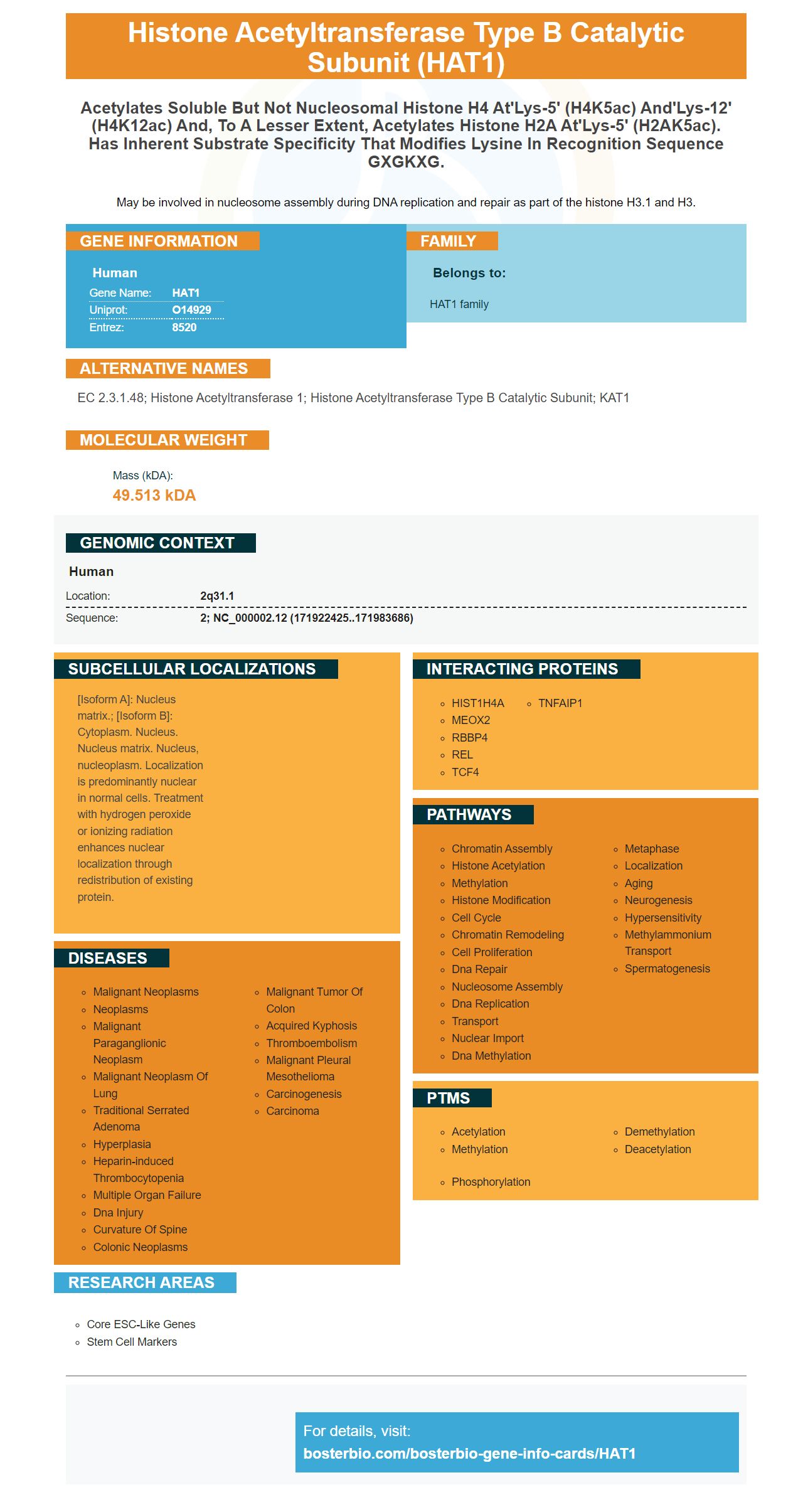
Facts about Histone acetyltransferase type B catalytic subunit.
May be involved in nucleosome assembly during DNA replication and repair as part of the histone H3.1 and H3.
Human | |
---|---|
Gene Name: | HAT1 |
Uniprot: | O14929 |
Entrez: | 8520 |
Belongs to: |
---|
HAT1 family |
EC 2.3.1.48; histone acetyltransferase 1; histone acetyltransferase type B catalytic subunit; KAT1
Mass (kDA):
49.513 kDA
Human | |
---|---|
Location: | 2q31.1 |
Sequence: | 2; NC_000002.12 (171922425..171983686) |
[Isoform A]: Nucleus matrix.; [Isoform B]: Cytoplasm. Nucleus. Nucleus matrix. Nucleus, nucleoplasm. Localization is predominantly nuclear in normal cells. Treatment with hydrogen peroxide or ionizing radiation enhances nuclear localization through redistribution of existing protein.
Boster Bio A03596-1 anti-HAT1 antibody is the best choice. This antibody reacts to Human and has been tested in ChIP and IF, IHC and WB. We will discuss the best uses for HAT1 and its antibody in this article. It is also possible to learn how bone marrow anti HAT1 antibodies can be used to perform a variety if research tasks.
The Anti-HAT1 Marker in Bostern Bio was developed to detect HAT1. This enzyme is an essential player in many biological processes, such as DNA replication, DNA repair, nucleosome assembly, and DNA repair. The boster bio antibody was created to recognize HAT1 monoclonally. It was validated in Western Blotting, Immunohistochemistry, and ELISA. The Boster Bio Anti-HAT1Marker is available in both monoclonal as well as polyclonal versions.
HAT1 interacts to the cenpAp46/48LIN53 Protein to inhibit the deposition CENP AHCP-3 on ACs. It interacts also with other proteins such as condensin II, the histone chaperone (RbAp46/48LIN-53) and condensin 2. These molecules are located in the nucleus and play an important role in chromatinization.
The physical interaction between HAT-1, CENP-AHCP-3 and HAT-1 is critical for segregation of nascent ACs. The RbAp46/48LIN53 Physical Interceptor also shares HAT-1 targets. The depletion of MYS-1 and HDA-1 had additive but not detrimental effects on AC segregation. The anti–HAT1 marker did no harm to HDA-1 and SET-1.
Boster Bio high affinity antibodies are a good choice for scientists who require primary antibodies for a variety of experiments. They are low in cross-reactivity and can be used to target specific proteins. These antibodies are monoclonal (or polyclonal) and have been widely used in research for more 25 years.
Boster is an expert in rabbit monoclonal antibodies with exceptional specificity, affinity and purity. Its PCD platform is designed specifically for target proteins in the Diagnostics & Therapeutics industry. This proprietary technology enables scientists to screen individual cells with proprietary chemistry that prevents B cells releasing antibodies and keeps them on cell surfaces. Plasma cells and splenocytes are incubated with flurochrome-conjugated antigens in order to identify the most specific antibody.
ELISAs have similar principles to other immunoassay technologies. They use specific antibodies to bind target antigens and a detection system to determine if the target antigen is bound to the target. Boster Bio has mastered the art of high-affinity prima antibodies. This increases the assay's sensitiveness. Scientists from all over the world rely on Boster Bio high-affinity primary antibodies.
Scientists must first create immunoglobulins in the host immune system before they can develop polyclonal primary antibody. The building blocks of primary antibodies are called complementarity-determining regions, and they are complex. Read more about complementarity-determining regions on Wikipedia. Then, they immunize and extract antibodies from the sera and eggs of the animals. Once the immune system recognizes the target protein, scientists can purify the protein and measure it.
Researchers can combine primary and second antibodies to answer more questions about specimens. This results in more context data and better answers. Boster Bio offers secondary antibody conjugates for a variety of applications. These high-affinity antibodies perform dual functions unlike other types. The primary antibody is used to detect the presence of target proteins, while the secondary one is used to recognize the protein that binds to the antigen.
When a primary antibody is recombinant, the genetic information of the antibody is preserved in a new protein, which is the target protein. This ensures that your research will be as accurate as possible. The supernatant can then be screened for positive or negative clones. You will receive an ELISA result, a map and 100ng of heavy strand plasmid plasmid genetic DNA.
The HAT1 gene plays a crucial role in bone marrow blood cell production. It is responsible the production of platelets, which aid blood to clot and fight infections. Most granulocytes will die within seven hours after being released. Half of them will survive in tissue for several days before returning into the bloodstream. Neutrophils, which attack bacteria and viruses, are the most common type.
Matching patients according to their tissue type is possible, although this may not be genetic. This is done using a comparison of the proteins in the donor's blood to those in the recipient’s bone marrow. A matching tissue type improves the likelihood of a successful transplant. The World Marrow Donor Association, (WMDA), is a network of hematopoietic tissue donor registries in 55 countries. WMDA lists 37.9 million potential donors, as well as 82,000 cord blood units. The NMDP is a great place to start your search for a donor.
In addition to its use in cell culture, the HAT1 gene is also useful for screening patients' marrow for cancer and other diseases. A fundamental process is molecular characterisation of a manufactured cell-batch. This article discusses the surface and molecular markers of MSC potency. HAT1 expression in bone-marrow MSCs is what makes them unique. qPCR analysis is an important first step in identifying candidates for treatment or research.
Patients should have a complete exam of their teeth before they undergo a bone marrow donation. This will help to reduce the risk for infection. The procedure may also involve a biopsy or bone-marrow aspiration. Bone-marrow harvesting is an easy procedure. Healthcare professionals often take bone marrow out of the posterior iliac, the sternum, and the upper tibia. However, the red bone marrow may be found in the upper portion of your tibia.
A bone marrow donation is the best way to fight cancer. A bone marrow donation from a relative is the most common option for patients. Only 30% of patients receive bone marrow from family members. 70% rely upon a bonemarrow donor from another person. High-dose chemotherapy and radiation can damage healthy stem cells during chemotherapy. This is why these transplants work so well. Diagnostics using bone marrow testing are useful for diagnosing certain conditions. They also help to assess iron levels, blood production, and other factors.
The HAT1 gene is a key regulator of the production of lymphocytes in the bone marrow. It helps to fight infection by recognizing invaders and sending signals to attack them. Adults have approximately 2.6kg red bone marrow. The highest concentrations of it are found in the marrow of the vertebrae. Bone marrow also produces monocytes, which are small blood cells that have a short lifespan in the blood. These cells mature into macrophages.
Healthcare professionals receive bone marrow donated by a donor who matches the patient’s HAT1 gene in a PBSC. Most PBSC donors go through the procedure in one session. Approximately 10% require two sessions. The sessions last between 2 and 4 hours. Unlike bone marrow transplants where anesthesia is required, PBSC donor does not require side effects. Most side effects subside within 7-10 days.
PMID: 9427644 by Verreault A., et al. Nucleosomal DNA regulates the core-histone-binding subunit of the human Hat1 acetyltransferase.
PMID: 11585814 by Makowski A.M., et al. Effects of acetylation of histone H4 at lysines 8 and 16 on activity of the Hat1 histone acetyltransferase.