This website uses cookies to ensure you get the best experience on our website.
- Table of Contents
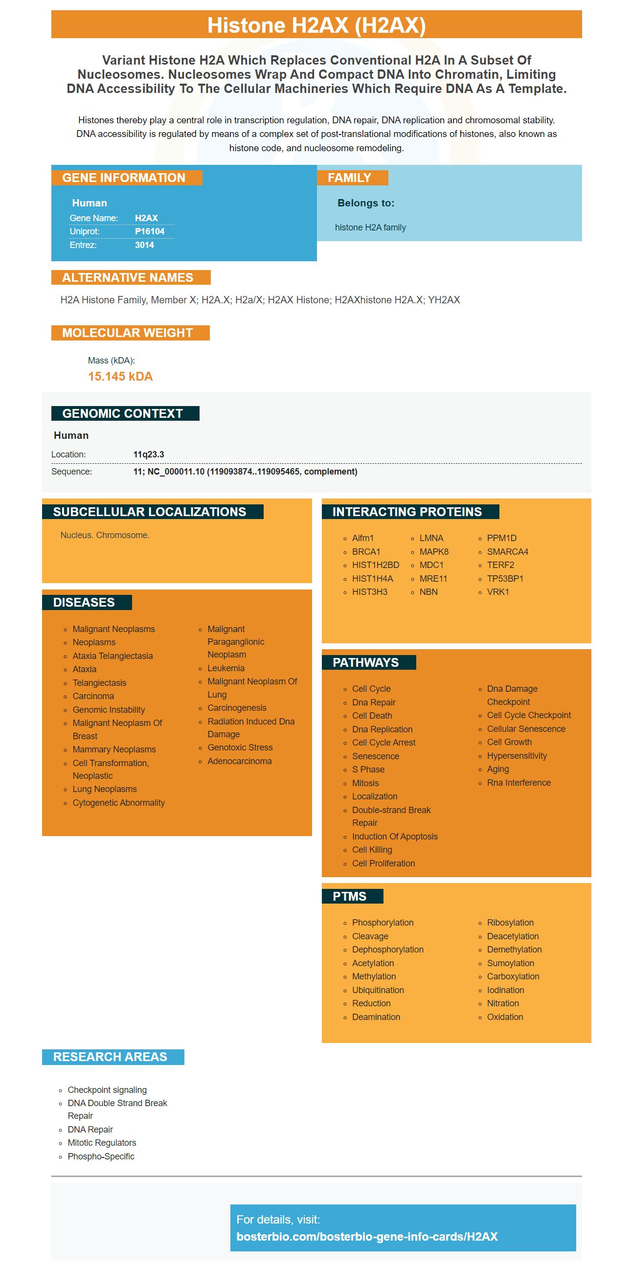
Facts about Histone H2AX.
Histones thereby play a central role in transcription regulation, DNA repair, DNA replication and chromosomal stability. DNA accessibility is regulated by means of a complex set of post-translational modifications of histones, also known as histone code, and nucleosome remodeling.
Human | |
---|---|
Gene Name: | H2AX |
Uniprot: | P16104 |
Entrez: | 3014 |
Belongs to: |
---|
histone H2A family |
H2A histone family, member X; H2A.X; H2a/x; H2AX histone; H2AXhistone H2A.x; YH2AX
Mass (kDA):
15.145 kDA
Human | |
---|---|
Location: | 11q23.3 |
Sequence: | 11; NC_000011.10 (119093874..119095465, complement) |
Nucleus. Chromosome.
Boster is universally useful for scientists all over the globe. No species, application, or sample type is excluded from its use. It is open to all researchers, as it does not have any validated results. It is important to know that this product has not been validated for specific species. This does not necessarily mean that the Boster product will not work for you.
gH2AX is a DNA DSB detection technique that has a high sensitivity. This technique is highly specific and works well for detecting DNA damage in testicles and bone marrow cells of mice. All cells produce the gH2AX proteins, including apoptotic and eukaryotic cells. This can influence their quantification.
This DNA damage detection method can detect low-level DSBs. The secondary antibody lanthanide-coupled to the DNA allows for significant detection of these mutations. The number of DNA DSBs correlated with an increase in fluorescence intensity. Time-resolved Fluorescence further enhanced detection of low level DNA DSBs. This method can also detect low-level DNA DSBs, since cells are able to absorb a higher dose of etoposide.
Using gH2AX to identify DNA DSBs is a better option than other methods. First, it is highly sensitive, and unlike the physical DSB, it registers cellular metabolic activities that help repair the DSB. The mechanism behind g-H2AX is related to DSB recovery. Moreover, gH2AX foci form after the IR.
Secondly, this method can detect the presence of DNA DSBs in a large volume of blood cells. Researchers can identify DNA double-strand breakage by analysing the gH2AX protein. These DNA damage lesions can seriously compromise the genome stability as well as cell survival. Double-strand breaks in DNA also initiate phosphorylation on Serine 139 the histone H2AX enzyme. This event requires activation DNA-PKcs ATM, ATR and DNA-PKcs which act as beacons for DSBs.
A fluorescent-tagged protein can also be used to detect DNA DSB. These fluorescent proteins, also known by nanobodies can be used to detect repair foci in living cells. This method allows researchers the opportunity to understand the mechanism of DSBs, and the role of repair in DNA. They can also track DNA DSBs and the phosphorylation gH2AX within the vicinity of DSBs.
Gamma H2AX (a phosphorylated histone H2AX) is a marker for DNA destruction. H2AX can be found in nucleosomes as part of the histone complex. It accounts for approximately two percent total H2A protein. Its phosphorylation, which is the first step in recruiting DNA-repairing proteins, is an excellent biomarker of damage.
Gamma H2AX can be detected by immunofluorescence or western blotting, and it serves as a biomarker for DSB frequency. GammaH2AX is a complex combination of multiple repair proteins. These include ATX mutated breast cancer, Nijmegen brokenage syndrome 1, NBS1, and Nijmegen breaking syndrome 1. It is an excellent target for drug development, in addition to its diagnostic utility.
Histone H2AX (a protein whose Phosphorylation can indicate a potential ovarian carcinoma relapse) is an example. It could eventually become routinely checked by medical professionals. Its high level is associated with higher tumor sizes, grade, and lymphode involvement. The clinical significance of this gene for breast cancer was examined by researchers. Gamma H2AX levels differed depending on the International Federation of Gynecology and Obstetrics(IFGO) stage. This suggests that this biomarker may prove useful in predicting relapse.
Healthy volunteers were injected with 900ml PBS pH 7.4 solution and then press onto their lower arms. The device was then secured with a tubular net wrap. The micro-arrays of the patches were used to measure biomarker concentrations. The biomarker was the Boster Bio H2AX solution.
The H2AX gene product g-H2AX is widely used to monitor drug and irradiations responses in cancer patients. The g-H2AX test can be used for both tumour biopsies as well as peripheral blood lymphocytes. For the assay, cells can be obtained by using mouthwash or by scraping the inner corners with a swab. Samples from the oral mucosa are not suitable for gH2AX analysis because they may contain dead cells, enucleated cell fragments, or squamous cells.
To determine radiosensitivity for patients who are considering radiation therapy, the gH2AX assay can be used. Radiation therapy has been proven to be one of most effective treatments for cancer. However radiation therapy can also cause severe side effects from normal tissue toxicity. Zwicker et. al. found that gH2AX measurements showed a linear relationship to radiation dose, while exposure to lymphocytes in patients who received 3D conformal therapy was the same.
A gH2AX focus is an indication that tumor cells have DSB repair defects. DSBs are often a sign that there is a tumor-related genetic impairment in patients with cancer. They are difficult to detect in vivo because of their low numbers. Scientists have recently discovered a way to detect gH2AX in vivo.
It is easy and cheap to monitor radiation and drug responses in patients by using g-H2AX. The assay works with a small biological specimen for quantitative analysis and can be automated. This biomarker can be used to measure radiation sensitivity and g-H2AX.
The gH2AX is a widely used marker in clinical studies for evaluating DNA damage and dose standardization. It has been used in various cancer types such as head and neck, breast, cervix, and adrenal renal. This biomarker could also be used in drug discovery and dosage optimization.
The gH2AX assay uses immunological techniques. It measures antibodies against the C-terminal of H2AX phosphorylated proteome in cell and tissue. The enzyme-linked immunosorbent test (ELISA), which measures gH2AX total concentration in tumor samples, lysates, and cells, is highly sensitive.
PMID: 2587254 by Mannironi C., et al. H2A.X. a histone isoprotein with a conserved C-terminal sequence, is encoded by a novel mRNA with both DNA replication type and polyA 3' processing signals.
PMID: 9488723 by Rogakou E.P., et al. DNA double-stranded breaks induce histone H2AX phosphorylation on serine 139.